by Jason Nenzel | Mar 27, 2025 | news
Reclaiming Your Life After a Herniated Disc: An Evidence-Based Approach to Recovery
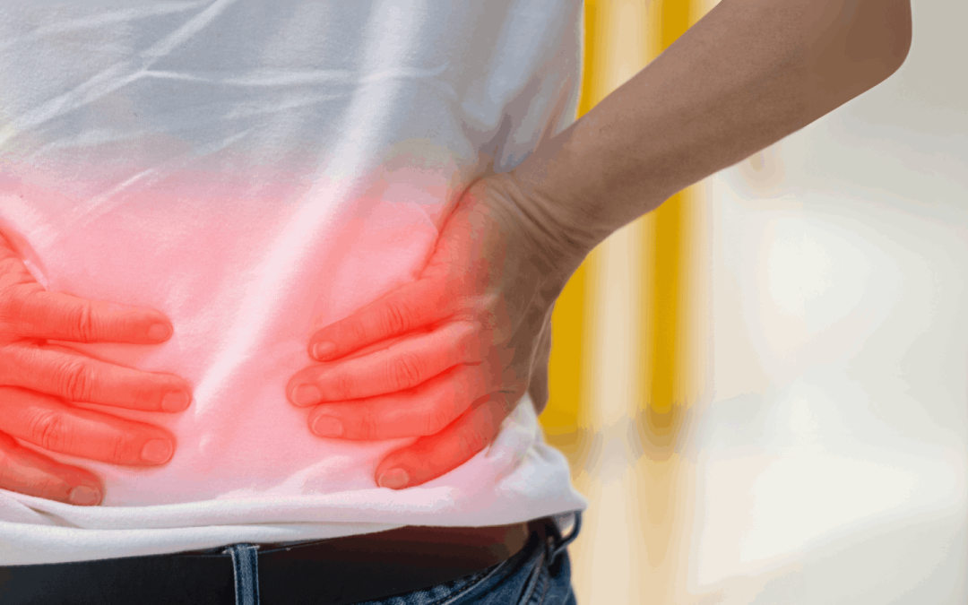
A herniated disc can be a life-altering condition, causing pain, limited mobility, and frustration. However, it is important to remember that the human body is highly adaptable, and with the right approach, most people can fully recover without the need for surgery. This article will explore the best evidence-based methods for reclaiming your life after a herniated disc, emphasizing conservative management and the body’s innate ability to heal.
Understanding Herniated Discs
A herniated disc, also referred to in lamen’s terms at times as a slipped or bulging disk, occurs when the soft, gel-like center of an intervertebral disc pushes through a tear in the tough outer layer. This can put pressure on nearby nerves, leading to symptoms such as lower back pain, leg pain (sciatica), numbness, and weakness. Herniated discs most commonly occur in the lumbar spine (lower back), but they can also develop in the cervical spine (neck), causing neck pain and discomfort radiating into the arms.
Causes of a Herniated Disc
– Wear and Tear: As we age, the intervertebral discs lose hydration and elasticity, making them less tolerant to physical stress.
– Sudden Strain: Lifting heavy objects improperly or engaging in abrupt twisting motions can cause a disc to herniate.
– Repetitive Stress: Jobs or activities that involve repetitive bending, lifting, or twisting can increase the risk of a slipped disk.
– Genetics: Some individuals may have a genetic predisposition to weaker discs, making them more susceptible to injury.
– Sedentary Lifestyle: Lack of movement and prolonged sitting can contribute to the weakening of spinal structures, increasing the likelihood of a bulging disk.
Conservative Management: The First Line of Defense
While surgery is sometimes necessary for severe cases, the vast majority of people with a herniated disc can recover through conservative treatments. Research consistently shows that non-surgical approaches are effective for managing symptoms and promoting healing.
1. Physical Therapy: The Backbone of Recovery
Physical therapy is one of the most effective ways to manage a herniated disk. A structured rehabilitation program can strengthen the muscles that support the spine, improve mobility, and reduce pain. A systematic review published in the Journal of Orthopaedic & Sports Physical Therapy found that targeted exercise programs significantly improve outcomes for individuals with lumbar disc herniation (Deyo et al., 2021).
Common exercises include:
– Resistance training: Strengthening the core muscles, improving overall fitness and understanding how to properly brace for lifting all reduces stress on the spine and supports recovery.
– McKenzie Method: A series of movements designed to alleviate pressure on the spinal nerves and promote healing.
– Mobility Exercises: Exploring the natural and complete range of lower back and hamstrings can relieve tension and improve mobility.
2. Medication for Pain Management
While medication does not heal a herniated disc, it can provide relief during the recovery process. Common medications include:
– NSAIDs (Non-Steroidal Anti-Inflammatory Drugs): Reduce inflammation and alleviate pain.
– Muscle Relaxants: Help manage muscle spasms associated with nerve compression.
– Oral Corticosteroids: Sometimes prescribed for short-term use to reduce severe inflammation.
According to a study published in the New England Journal of Medicine, NSAIDs have been shown to be effective in reducing lower back pain associated with disk herniation (Frymoyer et al., 2020). Although caution is recommended as prolonged use greater than 4 weeks can increase the risk of a GI bleed.
3. Spinal Manipulation: A Complementary Approach
Chiropractic care and spinal manipulation have been found to provide moderate relief for individuals with lower back pain due to disk herniation. A systematic review in The Spine Journal concluded that spinal manipulation can improve mobility and reduce pain when used alongside physical therapy (Rubinstein et al., 2021).
The Body’s Natural Healing Process
The body possesses a remarkable ability to heal itself, even in cases of a herniated disc. A study on sciatica recovery found that:
– 33% of patients recover within two weeks with conservative treatment.
– 75% of patients see substantial improvement after three months.
– 90% experience significant relief within six months.
The key to a successful recovery is patience and adherence to a well-structured rehabilitation program.
Optimizing Recovery: Practical Steps
To enhance healing and regain function, individuals should incorporate the following strategies:
1. Stay Active
While rest is essential in the initial phase, prolonged inactivity can delay recovery. Gentle activities like walking, swimming, and low-impact exercises help maintain mobility and reduce stiffness.
2. Adopt an Ergonomic Lifestyle
Proper posture and ergonomics play a crucial role in spinal health. Strategies include:
– Using a lumbar-supporting chair.
– Avoiding prolonged sitting.
– Sleeping on a firm mattress to maintain spinal alignment.
3. Weight Management
Excess weight can put additional strain on the lower back, increasing the risk of disk herniation. Maintaining a healthy weight through a balanced diet and regular exercise can aid recovery and prevent future issues.
4. Mind-Body Practices
Stress can exacerbate muscle tension and pain. Mindfulness-based stress reduction (MBSR), yoga, and meditation have been shown to improve pain perception and overall well-being in individuals with chronic back pain.
When to Consider Further Intervention
Although conservative treatment is effective for most cases, there are situations where further medical intervention may be necessary:
– Persistent Pain: If symptoms do not improve after three to six months of conservative treatment, additional evaluation is warranted.
– Neurological Deficits: Severe numbness, weakness, or loss of bowel/bladder control requires immediate medical attention.
– MRI Findings: Imaging studies may be recommended to assess the severity of the disk herniation and guide treatment options.
Final Thoughts: Embracing a Positive Mindset
Recovering from a herniated disk is a journey, but it is one that most people can successfully navigate with the right approach. The body is remarkably resilient, and with a combination of physical therapy, lifestyle modifications, and patience, individuals can return to an active and fulfilling life.
By focusing on evidence-based treatment options, maintaining an optimistic outlook, and committing to long-term spinal health, reclaiming your life after a herniated disc is entirely possible.
If you’re struggling with a herniated disc, incorporating targeted exercises and physical therapy into your recovery plan can be one of the most effective steps toward reclaiming your life and reducing pain. Let the expert practitioners at South Island Physiotherapy in Victoria, BC, help you develop a personalized treatment program tailored to your specific needs. Our team of Registered Physical Therapists, Massage Therapists, Kinesiologists, and Chiropractors are dedicated to providing exceptional care and guiding you through your recovery journey. Don’t let a herniated disc hold you back any longer – contact South Island Physiotherapy today to schedule a consultation and take the first step towards a pain-free, active lifestyle

by Jason Nenzel | Mar 20, 2025 | news
Understanding Cervicogenic Headaches: Diagnosis and Treatment
Headaches are among the most common health complaints, but not all headaches are the same. While many people are familiar with migraines and tension-type headaches, fewer are aware of cervicogenic headaches—a type of secondary headache originating from the cervical spine. Fortunately, conservative treatments, including physical therapy, can offer significant relief and improve quality of life.
What Is a Cervicogenic Headache?
A cervicogenic headache is a type of headache that originates from dysfunction in the cervical spine (the neck). Unlike primary headache disorders such as migraine and tension-type headache, a cervicogenic headache is a secondary headache, meaning it results from an underlying issue rather than being the primary condition itself.
Pain from a cervicogenic headache is often unilateral (on one side) and may be aggravated by neck movements or sustained postures. Referred pain from the upper cervical spine can travel to the head, causing symptoms that mimic other headache types. Headache symptoms may also include dizziness, blurred vision, and sensitivity to light or sound.
Diagnosis of Cervicogenic Headache
The International Classification of Headache Disorders (ICHD-3) provides diagnostic criteria for cervicogenic headaches, helping distinguish them from migraine and tension-type headaches. A proper diagnosis of cervicogenic headache requires:
- Headache originating from the neck and spreading to the head
- Reduced range of motion in the cervical spine
- Reproduction of headache symptoms through specific neck movements or pressure applied to the upper cervical spine
- Absence of primary headache disorder symptoms such as aura or nausea
Because other serious conditions, such as a tumor or vascular issues, can also cause secondary headaches, a thorough assessment is essential. A physical therapist or healthcare provider may use a combination of clinical examination and imaging studies to diagnose cervicogenic headaches accurately.
How Physical Therapy Can Help
Physical therapy is a highly effective, evidence-based approach to treating cervicogenic headaches. Since this type of headache originates in the cervical spine, addressing neck dysfunction can significantly reduce symptoms and prevent recurrence. Here are some key physical therapy interventions:
1. Manual Therapy
Hands-on techniques such as joint mobilization and soft tissue release help improve mobility in the upper cervical spine, reducing stiffness and pain that contribute to headache symptoms.
2. Postural Correction
Forward head posture is a common contributor to cervicogenic headaches. A physical therapist can assess posture and provide corrective exercises to restore proper cervical alignment, reducing strain on the neck.
3. Strengthening Exercises
Strengthening the deep neck flexors and upper back muscles improves overall neck stability, reducing excessive load on the cervical spine. This helps prevent headache recurrence and improves functional movement.
4. Range of Motion Exercises
Limited neck mobility is a hallmark of cervicogenic headaches. Targeted range of motion exercises help restore flexibility and reduce stiffness, allowing for pain-free movement.
5. Ergonomic and Lifestyle Modifications
Many daily activities, such as prolonged sitting and poor workstation setup, contribute to neck pain and headaches. A physical therapist can provide guidance on ergonomic improvements, sleep positioning, and activity modifications to reduce strain on the cervical spine.
Other Conservative Treatments
In addition to physical therapy, other conservative approaches can help manage cervicogenic headaches effectively:
- Heat and Cold Therapy: Applying heat can relax tight muscles, while cold packs may help reduce inflammation and pain.
- Massage Therapy: Soft tissue mobilization can relieve tension in the neck and shoulders, decreasing headache frequency.
- Acupuncture: Some individuals find relief through acupuncture, which targets trigger points that contribute to referred pain.
- Nerve Block Injections: In certain cases, a healthcare provider may use a nerve block to confirm the diagnosis and provide temporary relief by numbing the affected cervical nerves.
The Importance of Early Diagnosis and Treatment
Ignoring cervicogenic headaches can lead to chronic pain and decreased quality of life. Seeking early intervention from a physical therapist or healthcare provider ensures that the root cause is addressed, preventing long-term complications. Since cervicogenic headaches are a type of secondary headache, identifying and treating the underlying cervical spine dysfunction is key to lasting relief.
When to Seek Medical Attention
While most headaches are benign, some symptoms warrant immediate medical evaluation. Seek urgent care if your headache:
- Is sudden and severe (“thunderclap” headache)
- Occurs after head trauma
- Is accompanied by neurological symptoms such as numbness, weakness, or slurred speech
- Is associated with fever, weight loss, or persistent nausea and vomiting
- Changes in pattern or intensity over time
Conclusion
Cervicogenic headaches can be frustrating and debilitating, but effective treatment options exist. Physical therapy offers a safe and evidence-based approach to relieving symptoms, improving mobility, and preventing recurrence. By addressing posture, strength, and mobility, individuals with cervicogenic headaches can achieve long-term relief and return to daily activities with confidence.
If you suspect your headaches may be cervicogenic, consult with a physical therapist or healthcare professional for an accurate diagnosis and personalized treatment plan. With the right interventions, you can take control of your headache disorder and experience lasting improvement.
At South Island Physio, our expert practitioners specialize in treating various types of headaches, including tension headaches, migraines, and sinus headaches. We offer comprehensive assessments, manual therapy techniques, and customized exercise programs to address the root cause of your headaches. Our team of Registered Physical Therapists, Massage Therapists, Kinesiologists, and Chiropractors are dedicated to providing exceptional care you can trust. Don’t wait for your symptoms to worsen – contact South Island Physio today to schedule a consultation and start your journey towards a headache-free life. Let us help you restore your neck function, improve your posture, and reduce the frequency and intensity of your headaches. Your path to relief begins with a single call – reach out now and take charge of your health!

by Jason Nenzel | Feb 20, 2025 | news
Suffering From Depression and Anxiety? Physical Activity Can Help!
Anxiety is one of the most prevalent mental health conditions, affecting millions of people worldwide. According to the Anxiety and Depression Association of America, anxiety disorders impact nearly 40 million adults in the U.S. alone. While medication and talk therapy are commonly used to treat anxiety, alternative treatments for anxiety, including physical exercise, have gained significant attention. Research shows that regular physical activity may significantly reduce symptoms of anxiety and stress while improving overall mental and physical health.
Exercise and Depression and Anxiety Relief – The Science
Exercise and physical activity offer a wide range of mental health benefits. A systematic review and meta-analysis of multiple studies indicate that people with anxiety disorders who engage in physical activity experience a noticeable reduction in anxiety symptoms. Physical exercise may improve mood, cognitive function, and stress management by triggering various physiological changes in the body. Exercise activates the release of endorphins, often referred to as the brain’s “feel-good” chemicals, which help you manage anxiety and stress more effectively. Additionally, aerobic exercise influences the production of neurotransmitters like serotonin and dopamine, which play a crucial role in mental health. These biochemical changes can ease symptoms of depression and anxiety, making regular exercise a powerful tool for those dealing with anxiety and panic disorders.
Effects of Physical Activity on Mental Health
The effects of exercise on mental health disorders, including anxiety and depression, have been extensively studied. A systematic review and meta-analysis found that individuals who exercise regularly experience lower levels of anxiety compared to those who are physically inactive. The prevalence of anxiety tends to be higher in individuals with sedentary lifestyles, and reducing sitting time by engaging in physical activity may help alleviate symptoms.
The physical benefits of exercise, such as improved cardiovascular health and reduced inflammation, contribute to overall well-being, which in turn can have a positive impact on mental health. Regular physical activity helps regulate the body's stress response, reducing the burden of anxiety disorders and improving resilience against stress and anxiety.
How Much Exercise is Necessary?
The Department of Health and Human Services and the Centers for Disease Control and Prevention recommend at least 150 minutes of moderate aerobic activity or 75 minutes of vigorous exercise per week. Small amounts of physical activity, such as taking the stairs instead of the elevator or engaging in short walks throughout the day, can also provide benefits. Even less intense exercise, such as yoga or stretching, can help ease symptoms of depression or anxiety.
Forms of Physical Activity That Reduce Anxiety
Engaging in various forms of physical activity may help reduce anxiety symptoms. Some of the most effective types of exercise include:
1. Aerobic Exercise
Activities such as running, cycling, swimming, and brisk walking can significantly reduce symptoms of anxiety and depression.
2. Strength Training
Weightlifting and resistance exercises have been shown to have positive effects on mental health, reducing stress and boosting confidence.
3. Yoga and Tai Chi
These mind-body exercises combine physical movement with controlled breathing, helping to lower trait anxiety and improve emotional regulation.
4. Team Sports or Group Exercise Programs
Participating in formal exercise programs or recreational sports can provide social support, which is beneficial for managing anxiety.
5. Everyday Physical Activity
Simple activities like taking a walk after meals or encouraging children after school to engage in active play can contribute to better mental health outcomes.
Starting a New Exercise Routine
For those dealing with depression and anxiety, starting a new exercise program can feel overwhelming. Here are some tips for starting and maintaining an exercise routine:
- Set Realistic Goals: Start with small, manageable goals, such as a 10-minute walk daily, before gradually increasing intensity and duration.
- Choose Enjoyable Activities: Find physical activities that you enjoy, whether it’s dancing, hiking, or swimming.
- Exercise Regularly: Consistency is key; aim to engage in regular physical activity at least three to five times a week.
- Mix It Up: Incorporate different exercises to keep things interesting and target various muscle groups.
- Seek Support: Exercising with a friend or joining a class can provide motivation and accountability.
- Listen to Your Body: It’s important to recognize when to push yourself and when to rest, especially if you have a pre-existing health condition.
Exercise and Mental Health Professionals
While exercise offers many mental health benefits, it is not a substitute for professional care. If you experience severe anxiety and panic symptoms, it’s essential to consult a healthcare professional or mental health professional. A combination of therapy, medication, and regular physical activity may be the most effective approach for managing symptoms of depression or anxiety. The Mayo Clinic and other reputable health organizations recommend incorporating exercise into treatment plans under the guidance of a mental health professional.
The role of exercise in mental health treatment cannot be overstated. Regular physical activity can help reduce anxiety, improve mood, and enhance overall well-being. Every step counts, whether through structured aerobic activity, strength training, or simply making small lifestyle changes like taking the stairs instead of the elevator. Exercise and physical activity may not only ease symptoms of depression and anxiety but also provide long-term resilience against stress.
If you’re dealing with anxiety, incorporating an exercise routine into your daily life may be one of the most effective steps to take toward better mental health. Let the expert practitioners at South Island Physiotherapy in Victoria, BC, help you develop an exercise plan to support your mental health. Contact us today to schedule a consultation.

by Jason Nenzel | Dec 29, 2024 | news
How Kinesiology For Chronic Pain Helps People Feel Better
Chronic pain is a type of pain that lasts for a long time, often for months or even years. It’s different from acute pain, like when you accidentally stub your toe or cut your finger, which goes away quickly. Chronic pain can make it hard to do everyday activities like walking, working, or even enjoying time with friends and family. Kinesiology for chronic pain offers good news—movement and exercise, guided by an expert kinesiologist, can help!
Kinesiology is the study of how the body moves. Experts in this field use exercise and physical activity to help people recover from injuries, improve their health, and reduce pain. Let’s explore how kinesiology plays a big role in helping people with chronic pain feel better and get back to their normal lives.
Why Exercise is Key for Persistent Pain
Studies, including randomized controlled trials (a fancy way of saying carefully designed experiments), show that exercise can help people with chronic pain feel better. Exercise doesn’t just make your muscles stronger—it can also reduce pain, improve your mood, and help you do daily activities more easily. For example, aerobic exercise like walking, cycling, or swimming can help with low back pain, one of the most common types of chronic pain. These activities increase your heart rate and improve your overall health, including your cardiovascular system (your heart and blood vessels). People who stick to an exercise program often notice less pain and better movement.
How Kinesiology Designs Exercise Programs
Kinesiologists design exercise interventions (special plans for movement and workouts) based on the needs of each person. These programs might include:
- Aerobic Exercise: Activities like jogging, swimming, or brisk walking that improve heart health and endurance.
- Resistance Exercise: Strength training with weights, resistance bands, or even your own body weight to build stronger muscles.
- Endurance Exercise: Exercises that help you stay active longer without getting tired quickly.
- Manual Therapy: Hands-on techniques like massage or stretching to reduce pain and improve movement.
For example, older adults with chronic pain may need gentle exercises to improve their quality of life and keep them moving. In contrast, someone with lower limb pain might focus on strengthening their legs and improving balance.
What Does Science Say About Exercise and Pain?
There’s a lot of research showing that exercise works! A systematic review and meta-analysis (a big study that combines results from many smaller studies) found that exercise helps reduce pain and improve daily life for people with chronic pain. These studies show that exercise can:
- Reduce Pain Intensity: People feel less pain after doing the right kinds of exercise regularly.
- Improve Activities of Daily Living: Everyday tasks like cooking, cleaning, or climbing stairs become easier.
- Boost Mental Health: Physical activity can reduce stress and make people feel happier.
One randomized clinical trial showed that adults with chronic pain who followed a guided exercise protocol had a significant difference in their pain levels compared to those who didn’t exercise. The control group (the group that didn’t do the exercise) didn’t improve as much.
Matching the Right Exercise to the Person
Not all exercises work the same for everyone. The type, intensity, and frequency of exercise matter. For example:
- People with low back pain might benefit from low-impact movements like swimming or yoga.
- Those with chronic pain from cardiovascular disease might need carefully planned aerobic exercises.
- Resistance training can help adults build strength to support painful joints.
- Gentle exercises for older adults can help maintain mobility and independence.
Kinesiologists pay attention to these details to create safe and effective exercise plans.
Pain Management Beyond Exercise
While exercise is powerful, it’s often combined with other treatments, like physical therapy, manual therapy, and even counselling, to help manage pain. Together, these approaches create a recovery pathway that guides patients back to health.
For example, someone with chronic pain might see a physical therapist, follow an exercise program at home, and receive advice on how to move safely during daily activities. These combined therapies aim to reduce pain and gradually improve their life.
The Bigger Picture: Why Movement Matters
Whether it’s a structured exercise training program or simple movements like stretching or walking, staying active is key. Movement helps:
- Increase blood flow to painful areas, which can speed up healing.
- Strengthen muscles and joints, making the body more resilient.
- Improve mental health, reducing feelings of stress and anxiety that often come with chronic pain.
A Brighter Future Through Movement
Chronic pain doesn’t have to take over your life. With the help of kinesiology and well-designed exercise interventions, people can find their way to less pain and more freedom. From randomized controlled trials to real-life success stories, the evidence is clear: exercise works. Whether you’re an older adult, a busy parent, or a student, movement can help you feel better and do more of what you love.
So, the next time you hear someone say, “Exercise may reduce pain,” know that science backs it up. Through small, steady steps, movement truly shapes recovery pathways for people with chronic pain.

by Jason Nenzel | Nov 30, 2024 | news
Relieve Hip Bursitis: Causes, Symptoms, and Treatments That Work
Bursitis of the hip is a common condition involving painful swelling in the hip joint, often stemming from inflammation of the fluid-filled sacs that cushion the bones, tendons, and muscles. These sacs, called bursae, reduce friction and make movements around the hip smooth and pain-free. When these bursae become inflamed, it leads to hip bursitis, causing pain and discomfort that can affect daily activities. This article covers symptoms and causes, diagnostic methods, and the most effective treatment plans.
What is Hip Bursitis?
Bursitis refers to the inflammation of the bursa—a small, fluid-filled sac that cushions and protects the joints. Around the hip, there are two primary types of bursitis: trochanteric bursitis and iliopectineal bursitis. Trochanteric bursitis is the more common type, involving swelling of the bursa located at the top of the thigh bone. This bursitis is painful, and symptoms include pain on the outer side of the hip that can radiate down the thigh. Bursitis may worsen with movement, especially with repetitive hip motions.
Causes of Hip Bursitis
Hip bursitis can be caused by various factors. Some common causes include:
- Overuse of the Hip: Repeated stress on the hip from activities like running, cycling, or prolonged walking can put pressure on the hip, leading to bursitis. Overuse is a frequent cause for people whose jobs or studies require repetitive hip movement.
- Injury or Trauma to the Hip: A fall, bump, or any hip injury can inflame the bursa. Even minor trauma, when frequent, can cause this form of hip pain.
- Prolonged Pressure on the Hip: Sitting or lying on one side, especially on a hard surface for long periods places direct pressure on the hip, which can irritate the bursa over time.
- Muscle Imbalances or Weakness: Weak hip muscles may cause extra strain on the bursa. When supporting muscles, like the glutes, aren’t strong enough, it can lead to bursitis due to inefficient movement.
- Medical Conditions: Certain hip conditions can make someone more likely to develop bursitis. Arthritis, gout, or metabolic conditions can lead to bursitis by increasing inflammation in and around the hip joint. Septic bursitis—an infection in the bursa—is less common but requires urgent medical attention.
Symptoms of Hip Bursitis
The main hip bursitis symptoms include:
- Pain in the Hip and Outer Thigh: Bursitis is a painful condition often marked by a sharp or aching sensation around the hip and thigh. Pain in the hip often worsens when performing activities that cause pain, such as walking or climbing stairs.
- Tenderness and Swelling: You may feel tenderness in the area around the hip, especially when lying on or pressing against the affected hip.
- Increased Pain with Movement: Activities involving the hip—like hip abduction or climbing—often cause the pain to intensify. Sometimes even routine movements put pressure on the hip and worsen the discomfort.
- Difficulty Sleeping on the Affected Side: Sleeping on the side with bursitis can increase pain, disrupting sleep.
If you’ve noticed symptoms for the first time or are still experiencing symptoms despite rest, it’s a good idea to see a healthcare provider. A provider will help you find the right treatment plan, including options to relieve pain and improve range of motion.
Diagnosing Hip Bursitis
To diagnose bursitis, a healthcare provider will conduct a physical exam, asking you about your symptoms and examining the area around your affected hip. They may press on the hip to locate areas of tenderness and assess your hip’s range of motion to see what triggers pain.
Imaging tests, such as an X-ray, ultrasound, or MRI, may sometimes be ordered to confirm the diagnosis and rule out other common hip conditions like fractures or arthritis.
Treatment for Hip Bursitis
Once hip bursitis has been diagnosed, there are several effective treatment options. Treating hip bursitis generally involves a combination of self-care, medication, physical therapy, and sometimes, injections or surgery.
1. Rest and Activity Modification
Avoiding activities that aggravate the pain, such as running or prolonged walking, can help reduce inflammation in the bursa in your hip. Simple changes, like sitting on cushioned surfaces or switching sides when lying down, can reduce pressure on the hip.
2. Cold Therapy
Applying an ice pack to the area can reduce pain and inflammation. Apply the ice for about 20 minutes at a time, several times a day, particularly after activities that increase symptoms.
3. Over-the-Counter Pain Relievers
Medications like nonsteroidal anti-inflammatory drugs (NSAIDs), including ibuprofen and naproxen, can reduce inflammation and relieve pain. These drugs are typically safe for short-term use, but it’s best to discuss long-term options with a healthcare provider.
4. Physical Therapy
Working with a physical therapist can be very helpful for hip bursitis. Physical therapists create tailored exercises to strengthen hip muscles, support the joint, and improve flexibility.
Key exercises include:
- Strengthening Exercises: For the gluteal and core muscles, which stabilize the hip.
- Stretching Exercises: To maintain flexibility in muscles and tendons around the hip.
- Range-of-Motion Exercises: Help restore movement in the hip without irritating the bursa.
Hip abduction and other carefully prescribed exercises can be particularly beneficial. A physical therapist can monitor your progress and adjust exercises as needed.
5. Corticosteroid Injections
If the pain is severe or persistent, treatment for bursitis may involve corticosteroid injections. This is injected directly into the hip bursa to reduce inflammation. In many cases, corticosteroids offer significant pain relief, but their use should be limited, as repeated injections can weaken surrounding tissues.
6. Surgery for Trochanteric Bursitis
Surgery for trochanteric bursitis is rare but may be necessary for those who don’t respond to other treatments. Surgical options might include removing the bursa (remove the bursa). Surgery is generally reserved for cases where chronic bursitis affects quality of life, and other treatments have been ineffective.
Preventing Hip Bursitis
To prevent bursitis or avoid recurrence, you can take some proactive steps:
- Strengthen Hip and Core Muscles: Exercises that strengthen the glutes and core help stabilize the hip joint, reducing strain on the bursa.
- Maintain Flexibility: Stretching exercises for the hip, such as those targeting the IT band, glutes, and hip flexors, can keep the hip flexible, decreasing the chance of overuse.
- Warm Up Before Activity: Warm-up exercises help prepare muscles and joints, making them less susceptible to injury.
- Progressive loading: Rapid build-up volume of a new repetitive task ( i.e. running) can also cause bursitis. If you haven’t done a repetitive task for a long time or are just starting out, give yourself time to accumulate training volume.
- Practice Good Posture and Form: Correct form during activities like running or lifting is essential to avoid overuse of the hip.
- Avoid Prolonged Pressure on One Side: Avoid sitting or lying in one position for long periods, which can put pressure on the hip bursa.
Living with Hip Bursitis
Living with bursitis may require a few lifestyle changes to manage pain and prevent future flare-ups. Following prescribed treatments and working with healthcare providers to develop an individualized plan can greatly reduce symptoms.
If you’re still experiencing symptoms despite treatment, or if pain returns frequently, consult your healthcare provider. Persistent or severe pain may indicate that bursitis may require a change in treatment strategy. Working closely with a healthcare provider to manage hip bursitis can help you move more comfortably, and in most cases, hip bursitis usually gets better with consistent, attentive care.
Manage and Treat Hip Bursitis for a Pain-Free, Active Lifestyle
Hip bursitis is a common, manageable condition that affects people across age groups, especially those with active lifestyles or occupations that involve repetitive movements. While bursitis is a painful condition, understanding its causes and symptoms can help you find relief through personalized treatment. By following preventive steps and adhering to prescribed exercises, most people recover fully and can resume their regular activities.
Managing and treating hip bursitis requires a balanced approach—rest, strengthening exercises, and, if needed, medical interventions. With effective treatment, supportive physical therapy, and preventive measures, recovery from hip bursitis is achievable for most people, allowing them to continue leading active, pain-free lives.
Take the first step towards a pain-free life! Discover personalized treatment plans to help you move better, feel stronger, and live your best life. CONTACT US NOW to get started!


