by Colin Beattie | Nov 24, 2025 | news
Chiropractic Solutions for Sciatica: An Evidence-Based Guide
Finding sciatica pain relief starts with understanding the condition itself, and sciatica is one of the most common causes of lower back pain, affecting millions of people each year. It is characterized by pain that may travel from the lower back into the hip and often down the leg, radiating along the sciatic nerve. People describe it as sharp, burning, aching, or electric.
When you’re struggling with sciatica, daily activities can feel overwhelming. Fortunately, chiropractic care is one of the most widely used non-invasive approaches for managing and preventing sciatica, offering strategies that work with the body’s natural mechanics. This article explores the causes of sciatica, how chiropractic treatment can help, and the research on the benefits of chiropractic care for long-term management.
Key Takeaways
- Sciatica is a condition characterized by pain that radiates along the sciatic nerve, often due to compression of the nerve in the lower back.
- Chiropractic care for sciatica focuses on addressing the root cause of the pain, relieving pressure on the sciatic nerve, and improving movement.
- Chiropractic adjustments, mobilization, and soft-tissue work are common chiropractic techniques for sciatica that may offer pain relief, improved mobility, and effective management.
- Many people experience relief from sciatica, especially when chiropractic treatment is paired with appropriate exercise, lifestyle strategies, and education.
- While chiropractic care offers a natural approach, persistent or worsening symptoms should be evaluated by a healthcare professional.
What Is Sciatica? Understanding Sciatic Nerve Pain
Sciatica occurs when there is pressure on the sciatic nerve, often due to:
- A herniated disc
- Lumbar joint irritation
- Muscle tightness deep in the hip
- Spinal stenosis
- Inflammation
This nerve compression can produce sciatica symptoms such as:
- Pain shooting down your leg
- Tingling or numbness
- Weakness in the affected side
- Persistent sciatic pain
- Pain is often worse with sitting, bending, or lifting
Because sciatica is characterized by radiating pain, understanding its root causes is essential for effective management.
How Chiropractic Care Can Help With Sciatica
Chiropractic care offers a natural, non-pharmacological way to treat sciatica by improving spinal alignment and reducing mechanical stress. A chiropractor assesses how the spine, pelvis, and surrounding tissues may be contributing to your pain, then uses targeted chiropractic approaches to improve movement and decrease irritation.
Chiropractic care addresses the root cause of the pain
The goal is not simply a short-term reduction in symptoms, but lasting change through restoring movement and reducing compression. Chiropractic techniques aim to relieve pressure on the sciatic nerve, which may help reduce radiating discomfort and improve overall mobility.
Common Chiropractic Techniques for Sciatica
While techniques vary based on individual needs, research-supported options may include:
- Chiropractic Adjustments
Chiropractic adjustments can help restore movement in restricted spinal joints. This may:
- Reduce nerve irritation
- Improve spinal mobility
- Help relieve sciatica pain
Adjustments are among the most recognized chiropractic techniques for sciatica and have been shown to reduce back pain, improve function, and help manage recurring symptoms.
- Mobilization & Flexion-Distraction Techniques
Gentle, rhythmic mobilization can create space in the spine, which may:
- Reduce pressure on the sciatic nerve
- Improve motion
- Decrease discomfort
Flexion-distraction is often used in chiropractic for herniated disc conditions.
- Soft Tissue Therapy
Addressing muscle tension helps reduce strain on the spine and pelvis, improving the environment around the nerve.
- Lifestyle & Exercise Guidance
Although chiropractors may not prescribe medical treatments, many include education on:
- Exercise to support the spine
- Activity modification
- Strategies to reduce the risk of sciatica flare-ups
Movement and strengthening play a significant role in the long-term management of sciatica.
Benefits of Chiropractic Care for Sciatica
Many people seek chiropractic care because it:
- Provides sciatica pain relief without medication
- Helps alleviate pain caused by joint or nerve irritation
- Improves mobility in the lower back and pelvis
- Supports long-term relief when combined with exercise
- Offers a holistic approach to managing sciatica
Chiropractic care can help not only with pain relief, but also with understanding the cause of the pain, giving patients tools to manage symptoms beyond the clinic.
A Holistic Approach to Managing Sciatica
The most effective sciatica plans often combine:
- Chiropractic sessions
- Strengthening and flexibility exercises
- Load management education
- Lifestyle habits that reduce the risk of sciatica flare-ups
This well-rounded approach supports not only relief from pain but also long-term improvement in function and mobility.
FAQ
- Can chiropractic care help with sciatica?
Many people report significant improvement with chiropractic care. Techniques that address joint stiffness and muscle tension may help relieve pressure on the sciatic nerve.
- What causes sciatica?
Sciatica is often due to nerve compression from a herniated disc, joint irritation, muscle tightness, or inflammation.
- Is chiropractic treatment safe?
Chiropractic care is generally considered safe for many people when performed by a licensed provider. Individuals with complex medical conditions should consult their healthcare team first.
- Will chiropractic care provide lasting relief?
Many people experience meaningful, lasting improvement, especially when chiropractic care is combined with appropriate exercise and lifestyle strategies.
- Should I see a chiropractor for sciatica pain that radiates down my leg?
If you’re dealing with sciatic pain, radiating nerve discomfort, or lower-back symptoms that persist, a chiropractor for sciatica may help identify contributing factors and provide strategies for relief.
If sciatic pain is affecting your work, sleep, or daily routine, you don’t have to navigate it alone. At South Island Physio, our team takes a personalized, hands-on approach to understand the root cause of your symptoms and help you move with confidence again. With expert guidance, targeted treatment, and a supportive care plan, you can find long-lasting relief. Reach out today to book an appointment and take the first step toward feeling better.

by Colin Beattie | Nov 21, 2025 | news
Understanding How Manual Therapy Helps Your Back
Back pain, whether it’s acute low back pain, chronic low back pain, or non-specific low back pain, is one of the most common causes of disability worldwide. Many people assume that manual therapies must be performed only by a clinician such as a physical therapist. While some manual therapy techniques require professional skill, several manual therapies you can do on your own may help reduce pain and improve mobility.
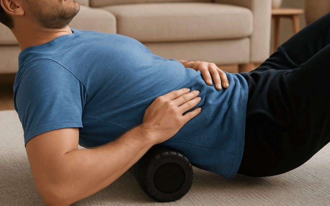
1. Self-Myofascial Release (SMR) With a Foam Roller or Ball
Self-myofascial release is a form of manual therapy commonly used for musculoskeletal pain and known as myofascial pain patterns.
Why It Helps:
Self-applied pressure can reduce pain, decrease muscle tone, and improve spinal mobility.
How to Do It:
– Place a ball or roller under your lower back or hips.
– Gently shift your weight to find a tender area.
– Hold 20–30 seconds and breathe.
2. Lumbar Self-Mobilization With a Towel Roll
A gentle self-version of lumbar mobilization.
Why It Helps:
Gentle pressure can improve movement and reduce discomfort.
How to Do It:
– Roll a towel and place it under your lower back.
– Rock your knees side to side or shift your pelvis.
3. Self-Massage Therapy for Paraspinal Muscles
Useful for acute and chronic low back discomfort.
Why It Helps:
Massage increases circulation and reduces muscle guarding.
How to Do It:
– Use your hands or a massage tool.
– Apply gentle pressure along the muscles beside the spine.
4. Self-Applied Trigger Point Therapy
Helpful for chronic musculoskeletal pain and chronic lower back pain.
How to Do It:
– Use a tennis or lacrosse ball.
– Press into tender knots for 15–45 seconds.
5. Self-Mobilizing Hip Flexor Release
Tight hip flexors can contribute to lower back pain.
How to Do It:
– Kneel on one knee.
– Lean forward until a stretch is felt.
– Add gentle pressure to the front of the hip.
Key Takeaways
- Several manual therapy techniques for back pain can be safely done at home and may help reduce pain and improve function, especially for patients with chronic low back pain.
- Techniques such as self-myofascial release, trigger-point pressure, and gentle mobilization often provide short-term relief from back pain and can support long-term recovery when combined with exercise.
- These self-techniques complement (but do not replace) care from a physical therapist as part of a complete treatment plan for treating back and neck pain.
- Consistent use may improve mobility, reduce stiffness, and help manage chronic musculoskeletal pain.
- If pain worsens, spreads, or involves more than muscle discomfort, professional evaluation is recommended.
FAQ
- Are these techniques safe for everyone?
Most people with nonspecific low back pain can use them, but red-flag symptoms require professional evaluation.
- How often should I do these techniques?
Daily or near-daily use is common.
- Will these techniques cure chronic pain?
They can reduce pain, but long-term improvement usually requires combining them with exercise and education.
- What if my pain returns?
Pain often returns without strengthening and movement retraining.
- When should I see a physical therapist?
If pain persists, worsens, or affects strength or sensation.
At South Island Physiotherapy, we know that every body is unique. Whether you’re seeking quick relief, long-term rehabilitation, or a balance of both, our team is here to guide you toward the care that best fits your needs. If you’re ready to understand the cause of your back pain and find a plan that works for you, book a consultation today. Together, we’ll design a personalized treatment strategy that supports lasting strength, mobility, and relief.

by Colin Beattie | Oct 28, 2025 | news
Finding the right care for your needs.
If you’re dealing with persistent neck pain, perhaps accompanied by shoulder pain, headache, or back and neck pain radiating down into your spine, you may find yourself asking, ‘should I see a chiropractor or a physiotherapist (physical therapist)?’ We’ll explore the approaches of chiropractic care and physiotherapy (physical therapy), look at what the evidence and clinical practice guidelines say, and help you decide the right treatment plan for you.
What is going on when you have neck pain?
Neck pain, or discomfort in the neck area, can arise from many causes. Poor posture (e.g., forward head carriage), sports injuries, stress, muscle tension in the upper back and neck, joint issues in the cervical spine (the neck portion of the spine), soft‑tissue strains, or chronic conditions all may play a role. The pain might be isolated to the neck area or accompany headaches, shoulder pain, or back pain. The cause of the pain helps guide the right treatment approach.
When you have neck pain, your range of motion may be reduced; you may feel joint pain, increased muscle tension in the neck and upper back, or even neck‑related dizziness in some cases. Treatment aims to reduce pain and inflammation, restore spinal alignment (especially if there’s a joint or spinal mobility component), improve soft tissue flexibility, improve range of motion, and help you manage pain (and prevent recurrence).
Two main healthcare professionals you might consider
Chiropractor (Chiropractic / Chiropractic Care):
A chiropractor typically focuses on the spine (and the neck area) and uses hands‑on techniques such as spinal manipulation (also called joint manipulation or spinal adjustment), joint mobilization, and sometimes soft tissue techniques or massage alongside the manipulation. The idea is to improve spinal alignment, reduce pain, improve range of motion, and relieve associated headache/back pain that comes from neck/upper‑spine dysfunction. In many clinics, chiropractors may use manipulation of the vertebral joints, often emphasizing the spine as the central system. Chiropractors focus on detecting and correcting what they call “subluxations” or misalignments (depending on the practitioner) and often apply joint manipulation to the spine and neck area.
Physiotherapist (Physiotherapy / Physical Therapy):
A physiotherapist addresses musculoskeletal impairments using a broader toolkit: exercise therapy (strength and endurance training, posture correction), manual therapy (which may include mobilizations, massage, soft tissue techniques), neuromuscular retraining for the neck/shoulder region, education (on ergonomics, posture, work habits), and functional rehabilitation. Physiotherapy tends to emphasize active treatments (you doing exercises) alongside passive treatments (therapist‐applied techniques) as part of a treatment plan that aims not only to relieve pain but to improve your overall functional ability and manage factors that may lead to recurrence.
So, in short, “chiropractor vs physiotherapist” is often a matter of emphasis: spinal manipulation and joint focus (chiro) vs broader functional rehabilitation and exercise focus (physio).
What do the clinical practice guidelines say?
For neck pain in general
A systematic review of clinical practice guidelines (CPGs) found that for non‑specific neck pain, radiculopathy, or whiplash‑associated disorders, the consistent recommendations were: assess for serious pathology, encourage activity, advise and reassure patients, and use exercise and manual therapies as part of management. Importantly, the guidelines emphasize that passive modalities alone (e.g., just manipulation, just massage) are not sufficient; a multimodal plan is preferred.
For example, the guideline “The Treatment of Neck Pain‑Associated Disorders and Whiplash‑Associated Disorders: A Clinical Practice Guideline” (2016) concluded, for recent‑onset (0‑3 months) neck pain (grades I‑II), the suggestion was to offer multimodal care; manipulation or mobilization; range‑of motion home exercise; or multimodal manual therapy. For persistent (>3 months) neck pain, the guideline suggests offering multimodal care or self‑management, and combining manipulation with soft tissue therapy, high‑dose massage, supervised group exercise, or home exercises.
For chiropractic treatment of neck pain
The guideline “Evidence‑based Treatment of Adult Neck Pain Not Due to Whiplash” (2005) focused on chiropractic populations and concluded that manipulation, mobilization, massage, strengthening exercises, and endurance training hold beneficial effects for chronic neck pain. More recently, “Best‑Practice Recommendations for Chiropractic Management of Adults with Neck Pain” (2019) found that for uncomplicated neck pain, including neck pain with headache or radicular symptoms, chiropractic manipulation and multimodal care are recommended.
For physiotherapy / physical therapy
Whilst there are fewer guidelines purely labelled “physiotherapy,” the general musculoskeletal neck pain guidance (see first item) emphasizes exercise (strength‑endurance, posture correction), manual therapy (mobilization, massage) in combination with active treatments. The evidence supports a physiotherapy role in exercise‐based rehabilitation, posture correction, and long‑term functional improvement.
Key takeaway from guidelines
- A multimodal treatment approach (exercise + manual therapy + advice/education) is widely recommended for neck pain.
- Manipulation or mobilization (spinal/neck) may have a role, especially when combined with other therapies.
- Exercise, posture correction, and active rehabilitation are essential. Physiotherapy excels here.
- For persistent or chronic neck pain, ongoing exercise, self‑management, and strength training become more important than just passive treatments.
- Screening for red flags (neurology, serious pathology) is critical.
What does the research say about effectiveness?
Effectiveness of physiotherapy for neck pain:
A systematic review examined physiotherapy interventions for chronic neck pain, finding that physiotherapy offers beneficial effects, particularly when incorporating strength and endurance training, multimodal physiotherapy (combining exercises and manual therapy), and massage/ manipulation/mobilization for chronic non-specific neck pain.
Effectiveness of spinal manipulation/chiropractic for neck and back pain:
Reviews of spinal manipulative therapy (SMT) – which is a core technique used by many chiropractors, show modest benefit for acute back pain in particular. For neck pain specifically, a 2003 review found that chiropractic spinal manipulation for neck pain did not convincingly show superiority over conventional exercise therapy. More recently, a systematic review focusing on spinal manipulative therapy for acute neck and lower back pain concluded that the evidence is limited and heterogeneous: SMT was associated with some improvements for acute neck pain, but there is considerable uncertainty.
Comparing chiropractic vs physiotherapy:
One randomized clinical trial in Sweden compared chiropractic and physiotherapy (for low back or neck pain) and found that both approaches, chiropractic and physiotherapy, as primary treatment reduced symptoms, and there was no significant difference in outcome or cost between the two groups after six months. Another more recent trial for chronic low back pain found no statistically significant differences in effectiveness or cost‑effectiveness between physiotherapy, chiropractic care, or combination treatment. (While this is back pain rather than neck, it gives a relevant indication.)
Safety / adverse outcomes:
A recent observational cohort study among older adults found that management of new onset neck pain with chiropractic manipulative therapy (CMT) was associated with lower rates of selected adverse outcomes compared to primary medical care with analgesic prescriptions. However, manipulation of the cervical spine is not without risk in rare cases; informed consent and proper screening are important.
Interpreting the evidence: What it means for you
Given the research and guidelines, here are key takeaways:
- Both physiotherapy and chiropractic care can reduce pain (neck pain and related back/neck issues) and improve function in many patients.
- Neither approach is uniformly “superior” in all cases: the Swedish RCT found little difference between chiropractic vs physiotherapy in terms of outcome or cost for neck/back problems.
- Physiotherapy has a stronger evidence base for exercise‑based rehabilitation, posture correction, strength/endurance training, soft tissue work, and long‑term functional improvement.
- Chiropractic (particularly manipulation/joint manipulation) may provide quicker relief in some cases of spinal/neck dysfunction, especially where joint restriction or spinal alignment is a contributor, but the evidence for superiority for neck pain specifically is weak.
- Safety appears acceptable for both when performed by qualified practitioners, but proper screening (especially for spinal/vascular issues) is necessary.
- Because neck pain often involves multiple contributing factor, e.g., poor posture, soft tissue stiffness, spinal joint dysfunction, weak neck/upper‑back musculature, a combined or integrative approach can sometimes be the best route.
Which one should you choose: Chiropractor or Physiotherapist?
Here are some guiding questions to help you decide:
- What is the cause of your neck pain?
If your discomfort is largely from muscle strain, poor posture, repetitive work habits, weakness in the neck/upper‐back, limited range of motion, then physiotherapy may be a strong choice: the focus will include strength and endurance training, posture correction, soft tissue work, and exercise to improve range of motion.
If you suspect joint dysfunction, stiffness in the cervical spine, or spinal misalignment contributing to your pain/headaches, or you’ve tried exercise without joint relief, seeing a chiropractor may help to address neck pain via spinal manipulation, joint mobilization, and spinal alignment.
- What do you want from treatment? Short‑term relief vs long‑term functional improvement?
If your priority is to reduce pain quickly, chiropractic care may offer hands‑on manipulation‑based relief.
If you’re focused on reducing the chance of recurrence, improving posture/work habits,and increasing strength and range of motion, physiotherapy offers a holistic approach to manage pain and improve function.
- What is your comfort level with joint manipulation and treatment style?
Some people prefer active engagement (doing exercises) and “learn how my body works” style, that aligns with physiotherapy. Others prefer hands-on adjustments and manipulation, as well as less self-driven rehab, which aligns with chiropractic care.
- Can you find integrative care or collaborative?
Many times physiotherapists and chiropractors may work in concert: chiropractic to relieve joint stiffness and increase range of motion, physiotherapy to build strength, endurance, posture control, and prevent recurrence. Asking for a treatment plan and agreement on goals is wise: e.g., manipulation or joint mobilization (by chiropractor) + exercise/soft tissue/massage (from physiotherapist) for a combined effect.
- Avoid red flags.
Whether you choose chiropractic or physiotherapy, if neck pain accompanies neurological symptoms (numbness, weakness in arms/hands, dizziness, neck‑related dizziness, signs of vertebral artery issues, trauma), you should first consult a healthcare professional (e.g., physician, neurologist) before manipulation.
Practical tips for treatment success
Ensure the treatment plan is clear: whether you’re seeing a chiropractor or physiotherapist, ask what techniques will be used (joint mobilization, spinal manipulation, massage techniques, exercise prescription) and what the goals are (reduce pain, improve range of motion, strengthen neck/upper back, correct weak posture).
Engage actively: even with chiropractic care, good outcomes often come when you also follow through with what you do between sessions (posture correction, home exercises, avoiding aggravating behaviours).
Focus on range of motion and soft tissue: gentle mobilizations, stretching tight upper‑back/neck muscles, improving mobility can help the spine and neck area move better, reducing muscle tension, and headaches.
Correct underlying contributors: poor posture (especially “tech‑neck” from phone/computer), weak neck/upper back muscles, sports injuries, repetitive strain — all should be addressed.
Be consistent: one session alone is rarely enough for chronic neck pain; a treatment plan over weeks (or months) with progressive exercise, manual therapy, manipulation/mobilization, and posture habits is typically needed.
Monitor outcomes: keep track of pain intensity, range of motion, function (e.g., ability to turn your head, do your sport/activity), headache frequency (if present), recurrence of back and neck pain. If things aren’t improving, reconsider the provider or approach.
Summary
In the debate of chiropractor vs physiotherapist for neck pain, the evidence suggests that both kinds of practitioners can help; neither is universally “best” in all cases. Physiotherapy has a stronger base for functional rehabilitation, strength/endurance and posture correction. Chiropractic care (via spinal manipulation and joint mobilization) may offer more immediate relief when joint/spinal alignment or stiffness is a significant contributor.
If you’re dealing with neck pain (with or without shoulder pain, headache, back pain) and you’re trying to decide whether to see a chiropractor or a physiotherapist (or both), the best choice depends on what is causing your neck pain, what you want from treatment, and how willing you are to engage in exercises/posture correction vs hands‑on adjustments.
A sensible approach: if joint stiffness or spinal alignment seems central (e.g., you feel your neck is “locked”, your range of motion is clearly restricted, manipulation has helped you in the past), then seeing a chiropractor could make sense. If your issue is more chronic, posture-related, muscle/soft tissue weakened, or you want to build long-term resilience, then physiotherapy might be the better starting point. And in many cases, a combined approach (chiropractic & physiotherapy care) can give you the best of both worlds.
Bottom line, whether to see a physiotherapist or a chiropractor isn’t an either/or decision if you find a provider who collaborates and provides evidence‐based care. The key is finding a healthcare professional you trust, who carries out an appropriate assessment, explains the treatment plan, tracks your progress, and works with you to reduce pain, improve your neck and back/spine health, restore your range of motion, and help you manage or prevent future episodes of neck discomfort.
At South Island Physiotherapy, we believe that real recovery from neck pain goes beyond short-term relief. It’s about restoring balance, building strength, and preventing pain from coming back. Our physiotherapists take a comprehensive approach, combining hands-on care, targeted exercises, and posture correction, to help you move confidently and comfortably again.
If you’re ready to understand the cause of your neck pain and find a plan that works for you, book a consultation today. Together, we’ll design a personalized treatment strategy that supports lasting strength, mobility, and relief.

by Colin Beattie | Oct 21, 2025 | news
Expert exercises and guidance for rehabilitation and back pain relief.
Physical Therapy and Rehabilitation play a crucial role in recovery from injury, surgery, or the wear-and-tear of a chronic condition, often demanding more than rest alone. For many people, an intentional exercise-based rehabilitation strategy under the guidance of a qualified clinician, such as a physical therapist, is central to restoring function, reducing pain, and returning to an active lifestyle. In this article, we’ll explore how rehabilitation exercises and a structured exercise program can support faster healing, and what you should know about technique, repetition, muscle control, and safe progression.
Why rehabilitation exercises matter
When a ligament, tendon, soft tissue or a major structure such as a joint is injured, the surrounding muscles that support the region often weaken, the range of motion becomes restricted, and pain can limit normal movement. For instance, in patients with low back pain, research shows that adding active exercise and physical therapy modalities leads to better improvement in pain and function than passive care alone. A broad review of musculoskeletal problems found that exercise was beneficial in many cases (though more research is required in specific sub‑groups).
By using targeted strengthening exercises, mobility work and controlled repetition, the goal is to restore muscle strength, neuromuscular coordination, and joint stability, so that tissues heal properly and you can safely return to your normal daily activities (like walking) or sport‑specific movements.
The role of the physical therapist in rehabilitation
A licensed physical therapist designs an individualized conditioning program (or part of a larger occupational therapy and rehabilitation plan) that uses evidence‑based methods. They assess your posture, movement patterns, muscle strength, flexibility and joint mechanics (for example, in the lumbar spine, shoulder blades, upper arm or whatever region is injured) and prescribe exercise progressions accordingly. According to guideline summaries for chronic pain, physical therapy is recommended when exercise is performed under supervision for 8‑12 weeks and functional improvement is present.
While self‑managed exercise programs are beneficial, they’re not a substitute for professional oversight when injuries involve complex structures (e.g., orthopaedic surgery, major soft tissue repair). The clinician ensures that your starting position is correct, that you perform the correct motion (such as flexion or extension) and that you avoid movements that might cause permanent damage or worsen the injury.
Key components of an effective rehabilitation exercise program
Let’s break down key principles and techniques that often appear in professional physical therapy and rehabilitation settings, especially for musculoskeletal issues like low back pain, shoulder pain or lower‑limb injuries.
1. Warm‑up & mobility
Every program ideally starts with a warm‑up to increase blood flow, heart rate and tissue temperature. Mobility and stability work often follows: for example, gentle movements that bring the joint through its available range of motion (ROM) without pain, helping prepare soft tissue, tendons, and ligaments for more intense loading.
A good warm‑up might include dynamic stretching, light aerobic movement (such as walking) or low‑load movement of the joint in multiple planes to improve readiness.
2. Strengthening and conditioning exercises
After warming up, the program will emphasize strengthening exercises to build or restore muscle strength and support the injured region. These may include:
- Isometric contractions: where you contract a muscle without a visible joint movement (for example, holding your core or glute tight for 10‑30 seconds). These are often used early when movement is painful or risky.
- Progressing to dynamic strengthening: performing movements (such as a squat, lunge, or single‑leg drop) through the full motion, returning to the starting position and repeating, using repetition to build endurance and strength of fast‑twitch and slow‑twitch muscle fibres.
- For the low back, research shows that exercise targeting trunk stabilizers, lumbar flexion/extension control, and hip‑muscle coordination helps improve outcomes.
- Exercises in other joints, such asthe shoulder (upper arm, shoulder blades) involve both abduction, adduction, internal and external rotation and scapular stabilization (for example, squeezing your shoulder blades, holding for a few seconds, then relax, repeat on the opposite side).
A well‑designed exercise program will also include general conditioning (cardio and endurance) to complement specific rehabilitation and improve overall health.
3. Functional movement and posture correction
Rehabilitation isn’t just about isolated muscle work; it’s about restoring function and ensuring you can move normally in daily life or sport. This means training movements like standing from a chair, walking gait, twisting, reaching overhead, lunge transitions and other functional tasks — under the eye of the care team, including your physical therapist and possibly an orthopaedic or sports medicine specialist.
For example, you might practise a controlled squat to teach correct hip/knee alignment, ensure the back muscles and glutes fire appropriately, and the spine remains neutral. In shoulder rehabilitation, you might begin with a scapular stabilization exercise (scapular retraction), then progress to more complex movements overhead or in a closed kinetic chain.
A change in posture often accompanies injury (e.g., guarding, stiffening). Rehabilitation exercises allow correction of this posture and help prevent compensatory movements that can lead to a chronic condition or pain elsewhere.
4. Range of motion, flexibility, soft tissue work
Once strength, mobility and stability are improving, rehabilitation will include stretching (feel a stretch in the muscle or adjoining tissues), soft tissue mobilization of tight tendons or ligaments and improving range of motion (for instance, hip flexion & extension, shoulder internal and external rotation). This is particularly important when scar tissue or stiffness limits motion.
For the low back, attention to hamstring tightness, hip flexor length, pelvic alignment and trunk mobility helps relieve load on lumbar discs and joints.
The starting position matters: you might begin lying on your back, knees bent to adjust the lumbar region, then perform controlled flexion/extension or rotation, returning to the starting position and repeat for a given number of reps.
5. Progression, repetition, monitoring
The professional technique emphasizes progressive overload: increasing the challenge, complexity or resistance of the exercise as you get stronger and more stable. Your physical therapist may prescribe you to perform a certain number of sets and repetitions for each exercise (for instance, 2‑3 sets of 10–15 repetitions of a lunge or single‑leg drop).
Monitoring progress is essential: Can you perform the movement without pain or with minimal soreness the next day? Are you improving muscle strength and joint stability? Are you ready to move to the next level (e.g., adding a dumbbell or increased resistance)?
In research, higher dosage (more practice) of specific task‑oriented exercise was shown to improve outcomes after stroke rehabilitation. While that’s a different population, the principle holds: repetition matters. Nevertheless, research on type, frequency and intensity of exercise for specific conditions (low back, shoulder, tendinopathy) remains less conclusive,
6. Return to function & prevention
Eventually, the goal is to restore function fully and enable an active lifestyle rather than just eliminating pain. Whether the issue was shoulder pain, a ligament or tendon repair, or low back dysfunction, you want to return to the activities you enjoy, such as sports, life tasks, work, recreation, all with confidence.
A conditioning program that integrates strengthening, flexibility, neuromuscular control and functional movement reduces the risk of re‑injury, helps prevent future episodes of pain and supports long‑term health. For instance, once you can safely perform a squat or lunge with good form, you may progress to sport‑specific or work‑specific tasks.
Working with a care team including your physical therapist, orthopaedics or sports medicine physician ensures that you follow safe criteria for return‑to‑sport (or return‑to‑activity). This is critical because prematurely returning can lead to setbacks or permanent damage.
Sample rehabilitation exercise for the lower back (illustrative only)
Here is a simplified sample sequence that illustrates some of the principles discussed. (Note: this is not a prescription, you should consult your physical therapist for your specific condition.)
- Starting position: Lie on your back with knees bent, feet flat on the floor, arms at your sides.
- Pelvic tilt: Gently flatten your lower back (lumbar) onto the floor by engaging your deep core and glutes. Hold for 5 seconds, relax to the starting position. Repeat 10 times.
- Bridging: From the same starting position, lie on your back, engage glutes and hamstrings and lift your hips off the floor until your body forms a straight line from shoulders to knees. Hold for 3‑5 seconds, then lower back to the starting position. Perform 2 sets of 10 repetitions.
- Quadruped opposite arm/leg raise (bird‑dog): On all fours (hands under shoulders, knees under hips), extend your right arm forward and left leg behind, keeping your back flat, then return to the starting position and repeat on the opposite side. Perform 10 repetitions on each side.
- Lunge forward: Stand and take a step forward into a lunge, keeping your back upright and your front knee over your ankle. Return to the starting position and repeat on the opposite side. Do 2 sets of 8–10 per leg.
- Squat: With feet shoulder‑width apart, bend your knees and hips as if sitting back into a chair, keeping your spine neutral and back muscles engaged. Lower to your comfort level (e.g., thighs parallel or slightly above), then push through your heels to return to the starting position. Do 3 sets of 8–12 repetitions.
Throughout this sequence, emphasis is on mobility and stability, controlled movement, pain‑free execution and correct posture.
Evidence & what it tells us … and what it doesn’t
The research base for therapeutic exercise and physical therapy in musculoskeletal conditions is growing:
- A review demonstrated exercise had positive effects on conditions like low back pain, fibromyalgia, incontinence and stroke rehabilitation.
- A randomized‑controlled trial in chronic non‑specific low back pain found that combining physical therapy modalities and exercise improved pain and function more than exercise and medical treatment alone.Guidelines for chronic pain management emphasize that exercise‑based physical therapy is recommended, although the exact frequency, type and duration need further study.
- For neurological rehab (stroke), evidence shows that task‑oriented training and repetition have a stronger effect than classic passive treatments. However, it’s important to recognize limitations: For many specific injuries (e.g., specific tendon repairs, ligament reconstructions, shoulder pathologies), the optimal type of exercise, progression rate and dosage are less well defined. In the domain of low back exercises, a review noted that “the scientific foundation to justify their choice is not as complete as one may think”.
In other words: exercise works, but it matters how you implement it (technique, supervision, progression) and ensure it fits your condition, goals and timeline.
Practical tips for a safer, more effective rehab exercise approach
- Always begin in the starting position that your physical therapist identifies — correct alignment, neutral spine, and safe movement path.
- Focus on repetition with good form rather than doing more reps poorly. It’s far better to perform 10 perfect squats than 30 sloppy ones.
- Progress gradually. When you can perform an exercise comfortably (e.g., 3 sets of 12 reps without pain or excessive soreness), then you might increase resistance (add a dumbbell), add a challenge (single‑leg variation, change in posture), or move into sport‑specific movements.
- Monitor pain. You should not provoke sharp or intense pain (though some mild discomfort or soreness can be acceptable). If you feel a sudden increase in pain, stop and communicate with your physical therapist or care team.
- Include both mobility and stability elements in your exercise program. A joint that moves well but lacks muscular control remains vulnerable; conversely, strength without mobility often leads to compensations.
- Make sure your program addresses the entire kinetic chain. For example, if you have shoulder pain or weakness in the upper arm or shoulder blades region, your rehab should include scapular stabilization and rotator‑cuff control, not just local shoulder flexion/extension.
- Integrate your rehabilitation exercises into your broader healthy lifestyle — i.e., combine them with general conditioning, cardiovascular fitness, good nutrition, and postural awareness.
- Stay engaged in your care team. Communication between your orthopaedic, sports medicine specialist (if applicable) and your physical therapist ensures that your rehabilitation aligns with any surgical or diagnostic issues and your return to the starting position criteria or return to activity timeline is safe.
- Recognize that prevention is also part of rehabilitation. Once you’ve restored function, maintain it. Regular strengthening and mobility work help prevent future episodes of pain, degenerative changes or chronic condition development.
Final thoughts
Rehabilitation through exercise under the supervision of a physical therapist is a powerful path to faster healing, restoration of function and prevention of future injury. Whether you are dealing with low back pain, shoulder discomfort, soft tissue injury of a tendon or ligament, or recovering from orthopaedic surgery, following a structured plan with correct exercise technique, appropriate repetition, and progression makes a real difference.
Studies support that combining exercise with professional physical therapy modalities leads to better outcomes than passive care alone. Although research is still refining the exact “best” approach for every condition, the consistent message is: Movement matters, repetition matters, and doing it well matters.
By actively engaging in your rehabilitation, committing to the program your physical therapist sets, monitoring your progress, and integrating your exercises into your daily life; you give yourself the best chance to heal faster, regain strength, move without pain and return to the activities you love.
At South Island Physiotherapy, we know that every body, and every injury, is unique. Whether you’re seeking quick relief, long-term rehabilitation, or a balance of both, our team is here to guide you toward the care that best fits your needs. By understanding the differences between physical therapy and rehabilitation, you can make an informed choice, and we’ll be here to help you move with confidence every step of the way. Contact South Island Physio today!

by Colin Beattie | Sep 22, 2025 | news
Chiropractic vs Physiotherapy: Understanding the Differences and Choosing What’s Right for You
When dealing with back pain, neck pain, or joint pain, many people wonder whether to see a physiotherapist or chiropractor. Both disciplines provide hands-on care for disorders of the musculoskeletal system, but they approach pain relief, rehabilitation, and long-term physical function in different ways. This article explores the similarities and key differences between chiropractic and physiotherapy, so you can make an informed decision about which healthcare provider is best for your specific health condition.
What Physiotherapy Focuses On
Physiotherapy (also known as physical therapy) focuses on restoring movement, function, and quality of life. A physiotherapist is trained to assess a wide range of conditions, from sports injuries and knee pain to lower back pain, post-surgical recovery, and neurological conditions.
A physiotherapist will more commonly use:
- Exercise and mobility training
- Soft tissue techniques and manual therapy
- Education and training about posture, ergonomics, and load management
- Progressive physiotherapy treatment plans to improve strength, flexibility, and confidence in movement
In most cases, physiotherapy sessions aim not only to decrease pain but also to address the root cause of the problem by improving overall movement capacity.
What Chiropractic Care Primarily Focuses On
Chiropractic care is a discipline that emphasizes spinal manipulation and other adjustments and manipulations of the musculoskeletal system, particularly the spine. A doctor of chiropractic (chiropractor) is trained to evaluate mechanical disorders of the musculoskeletal system and provide chiropractic services aimed at restoring range of motion and reducing pain and stiffness.
A chiropractor is recommended when patients want hands-on treatment to help with back and neck pain, headaches related to mechanical issues, or certain joint pain conditions.
Chiropractors use chiropractic treatment methods such as:
- Spinal manipulation and mobilization
- Manual therapy for joints and soft tissues
- Advice on exercise and lifestyle modifications
- Short-term treatment plans aimed at pain reduction
Chiropractors and Physiotherapists: A Lot of Overlap
Despite their differences, chiropractors and physiotherapists share many similarities. Both work within the realm of musculoskeletal issues, use hands-on methods like manual therapy, and aim to reduce pain, improve range of motion, and restore physical function. Both also help patients with pain and stiffness return to everyday activities and optimal health. This is why many people are unsure about the difference between physiotherapy and chiropractic or wonder which to choose for a specific health condition.
Key Differences Between Chiropractic and Physiotherapy
Here are the top 5 differences to keep in mind when comparing chiropractic vs physiotherapy:
- Treatment Style
– Chiropractic care primarily focuses on spinal manipulation and joint adjustments and manipulations.
– Physiotherapy focuses more on exercise-based rehabilitation, long-term education and training, and a variety of techniques to restore function.
- Treatment Plan Duration
– A chiropractor is recommended for shorter courses of chiropractic treatment aimed at pain relief and mobility.
– A physiotherapist may build a longer-term plan involving exercises to strengthen muscles and prevent recurrence.
- Education and Training
– A doctor of chiropractic completes chiropractic college, specializing in spinal manipulation and manual therapy.
– A master of physiotherapy program trains physiotherapists in a broad range of conditions, including post-surgical care, sports rehab, and neurological rehabilitation.
- Conditions Treated
– Chiropractic care vs physiotherapy differs in scope: chiropractic care primarily focuses on back and neck pain, while physiotherapy can also address knee pain, post-surgical care, and chronic conditions.
- Approach to Self-Management
– Physiotherapy treatment often emphasizes education, exercise, and prevention.
– Chiropractic services often emphasize hands-on care and manipulation for immediate symptom relief.
Frequently Asked Questions
- Should I see a chiropractor or physiotherapist for lower back pain?
Both can help. A physiotherapist would often design a program with exercises to strengthen your back, while a chiropractor is recommended if you want manual therapy or spinal manipulation to quickly relieve pain and stiffness.
- What’s the difference between chiropractic care and physiotherapy for sports injuries?
Physiotherapy and chiropractic can both treat sports injuries. Physiotherapy sessions often include rehabilitation exercises, while chiropractic services may use manual therapy and adjustments to restore mobility.
- Can physiotherapy and chiropractic care be combined?
Yes. Many patients benefit from a treatment plan that includes both chiropractic and physiotherapy at different stages of recovery, depending on their health condition.
Choosing What’s Right for You
When deciding between chiropractic vs physiotherapy, think about your goals. If you need immediate pain relief from back pain or neck pain, a chiropractor is recommended for spinal manipulation and short-term care. If you want a longer-term plan focused on rehabilitation, education, and exercises to strengthen and prevent recurrence, a physiotherapist will more commonly use the right strategies. For many musculoskeletal issues, combining both physiotherapy and chiropractic care may provide the best balance between quick symptom relief and lasting physical function.
Final Thoughts
Both chiropractic care and physiotherapy services are effective, evidence-based ways to reduce pain and improve movement without pain. The real difference between chiropractic care and physiotherapy lies in emphasis: chiropractors use chiropractic treatment methods like spinal manipulation for rapid relief, whereas physiotherapy focuses on long-term recovery through exercise, education, and rehabilitation. Understanding the differences between chiropractic and physiotherapy helps you choose the right therapist and treatment plan for your health condition—and ultimately return to life with less pain and greater confidence in your body.
At South Island Physiotherapy, we know that every body, and every injury, is unique. Whether you’re seeking quick relief, long-term rehabilitation, or a balance of both, our team is here to guide you toward the care that best fits your needs. By understanding the differences between chiropractic and physiotherapy, you can make an informed choice, and we’ll be here to help you move with confidence every step of the way. Contact South Island Physiotherapy today to schedule your next appointment.

by Colin Beattie | Sep 12, 2025 | news
Dry Needling vs. Acupuncture: Key Differences, Similarities, and When Each Works Best
If you’ve been dealing with musculoskeletal pain, chronic tightness, or stubborn trigger points, you’ve probably heard of dry needling vs acupuncture debates. Both therapies insert needles into specific areas of the body to promote relief from pain, improve range of motion, and enhance recovery. But while they look similar at first glance, the key differences between dry needling and acupuncture lie in their history, intent, and evidence base.
This post will explore the distinctions between dry needling and acupuncture, what the research says about each, and how to decide which may be right for your injury or condition.
What’s the Difference Between Dry Needling and Acupuncture?
At a glance, both methods use thin needles to stimulate the body—but their philosophies and goals are distinct.
- Dry needling is a modern Western medical technique.
- Dry needling is a technique often performed by physical and sports injury therapists or physiotherapists.
- It focuses on inserting filiform needles directly into myofascial trigger points—tight bands of muscle that contribute to pain, stiffness, or movement restriction.
- Dry needling treatment is part of a broader physical therapy approach, often combined with exercise and education.
- Acupuncture is based in traditional Chinese medicine.
- Acupuncture involves inserting acupuncture needles into meridians and energy points believed to influence the body’s flow of “qi.”
- In the West, the use of acupuncture has expanded, and it is now widely studied for pain management, chronic pain, and systemic conditions.
- An acupuncture session is performed by physical therapists in some states but most often by a licensed acupuncturist, certified by the National Certification Commission for Acupuncture and Oriental Medicine (NCCAOM) or similar bodies.
So, dry needling and acupuncture involve very similar needles used, but their intent differs: dry needling treats muscles and trigger points, while acupuncture treats musculoskeletal pain and other conditions through meridian theory.
Needling Techniques: How They Work
- In point dry needling, a needle is inserted directly into a trigger point, sometimes producing a local twitch response.
- Non-trigger point dry needling places needles into specific points around the painful region to restore movement and blood flow.
- Acupuncture and dry needling also differ in the type of needle: both use filiform needles, but acupuncture is performed with different patterns, depth, and locations depending on diagnosis within acupuncture and oriental medicine.
While both needles are used to stimulate tissue, acupuncture may aim to restore energetic balance, whereas dry needling is based on neuromuscular and biomechanical models.
The Evidence: Does It Work?
Evidence for Dry Needling
Research shows that:
- Dry needling can help reduce muscle tension and improve pain and movement issues associated with back pain, neck pain, and sports injuries.
- Meta-analyses suggest that dry needling is more effective than sham treatments for short-term pain relief and improving range of motion.
- However, the effectiveness of dry needling may depend on combining it with part of a broader physical rehabilitation plan.
The American Physical Therapy Association considers dry needling a safe and useful adjunct when performed by physical therapists who are trained and licensed in states that allow physical therapists to perform dry needling.
Evidence for Acupuncture
Studies show that:
- Acupuncture works for chronic pain, particularly acupuncture for osteoarthritis, migraines, and back pain.
- Clinical trials find benefits of acupuncture compared to sham acupuncture, though the effect sizes can be modest.
- The groups for acupuncture worldwide point to its role in reducing reliance on opioids and other drugs for pain management.
- Potential risks of acupuncture are low but may include bruising, dizziness, or infection if not properly sterilized.
Overall, both therapies provide measurable relief from pain, though high-quality studies suggest results are strongest when combined with movement-based rehabilitation.
Which Should You Choose?
When deciding between acupuncture or dry needling, consider your goals:
- Choose dry needling if you’re looking for direct treatment of trigger point-related muscle pain, tightness, or local movement restriction. A dry needling session is often performed by physical therapists and paired with rehab exercises. Dry needling may help athletes and those with sports injuries in particular.
- Choose acupuncture if you want a holistic approach to reduce pain, manage chronic pain, or address systemic concerns alongside pain and muscle tension. Acupuncture currently has broader recognition for conditions like migraines, stress, and acupuncture to reduce osteoarthritis symptoms.
Final Thoughts
So, dry needling vs acupuncture isn’t about which is “better,” but which fits your needs. If you’re looking for relief from pain and movement issues associated with sports injuries or musculoskeletal pain, dry needling is also a strong option—especially when used as part of a larger pain management plan with physical therapy.
Meanwhile, acupuncture is practiced by tens of thousands worldwide, and acupuncture may be the right choice if you prefer a whole-body approach rooted in traditional Chinese medicine.
Both therapies insert needles into specific sites, but their philosophies differ. By understanding the difference between dry needling and acupuncture, you can choose wisely—or even benefit from both.
At South Island Physiotherapy, we believe that pain relief should be more than temporary. It should set the stage for lasting strength and movement. Whether through dry needling, acupuncture, or a tailored rehabilitation plan, our goal is to help you recover, move freely, and feel your best.
If you’re curious about which treatment is right for you, book a consult with a physiotherapist today. Together, we’ll design a plan that supports your recovery and keeps you moving with confidence.


