
by Jason Nenzel | Sep 26, 2024 | news
Enhancing Patient Care with Essential Resources and Evaluation Techniques
In the world of physiotherapy, ensuring that treatments are both effective and personalized is essential. This is where evidence-based practice (EBP) comes into play. EBP is about making informed clinical decisions by combining research evidence, clinical expertise, and patient values. But what does it mean in the context of physiotherapy, and why should it matter to you?
In this blog, we’ll explore the concept of evidence-based practice in physiotherapy, its benefits, and how it impacts your care.
What is Evidence-Based Practice?
The concept of evidence-based practice was first introduced by Sackett DL and Richardson WS, pioneers in the field of evidence-based medicine. Their work laid the foundation for clinical practice that emphasizes the integration of best research evidence, clinical expertise, and patient preferences. Sackett’s approach is widely used today, shaping the practice of EBP in fields like physiotherapy.
Evidence-based practice in physiotherapy is an approach that integrates the best available evidence with the clinician’s expertise and the preferences of the patient. It’s a clinical decision-making process that ensures the physical therapist uses the most up-to-date, scientifically supported treatments.
Let’s break it down into its core components:
1. Best Available Evidence
At the heart of evidence based practice is the use of the best available evidence. Physiotherapists rely on systematic reviews, clinical trials, and other high-quality scientific evidence to guide treatment decisions. For example, if you’re dealing with a sports injury, the physiotherapist should rely on research that highlights the most effective clinical practice guidelines and rehabilitation techniques, backed by studies from sources like BMJ or systematic reviews in medical journals to inform their clinical decision making.
2. Clinical Expertise
While research is critical, clinical expertise plays a significant role in making effective decisions. Experienced clinicians know how to adapt evidence-based medicine (EBM) to individual values and circumstances. This expertise ensures that your treatment is personalized based on your condition, how you respond to therapy, and any specific needs you may have.
3. Patient Values and Preferences
In evidence-based physiotherapy, your input is just as important as the research and expertise of the clinician. You have the right to make decisions about your care, and EBP ensures that your values, goals, and preferences are always taken into account. This could mean choosing one type of therapy over another based on your lifestyle, comfort, or specific recovery goals. Ultimately, injury recovery is about collaboration not dictation and evidence-based physical therapy should seek to include patients throughout this process.
Why Evidence-Based Practice is Important in Physiotherapy
EBP is a cornerstone of modern physiotherapy because it provides many key benefits:
- Better Outcomes: By basing treatment on best available evidence, physiotherapists can deliver effective, research-backed interventions that result in improved rehabilitation and faster recovery and more predictable outcomes.
- Up-to-Date Care: Evidence-based physiotherapy ensures that clinicians stay current with the latest advancements in evidence-based medicine, using the most recent and trusted sources of evidence to guide treatment plans.
- Personalized Care: EBP combines scientific findings with your individual preferences, ensuring that treatments are tailored to your needs.
- Cost-Effective Treatments: Relying on evidence-based medicine helps avoid unnecessary or ineffective interventions, leading to more efficient and affordable care.
How Physiotherapists Use Evidence-Based Practice
Here’s how evidence-based practice is applied in real-world clinical practice:
- Asking Clinical Questions: Healthcare professionals who practice evidence based medicine, like those at South Island Physiotherapy, formulate questions in a manner that seeks to create clarity and understanding for both the patient and the clinician. This involves active listening, validation and humility on behalf of the therapist in order to formulate specific questions that guide their diagnostic process.
- Searching for Evidence: They rely on databases like BMJ, PubMed, and the Cochrane Library to find best available evidence and systematic reviews that are relevant to your condition.
- Evaluating the Evidence: Physiotherapists critically appraise the available evidence to ensure that it is reliable and applicable to your situation, often following practice guidelines set by professional bodies.
- Applying the Evidence: After assessing the evidence, clinicians integrate it with their clinical expertise and your preferences to create a treatment plan tailored specifically to you.
- Evaluating Outcomes: Once treatment begins, your physiotherapist will continuously monitor your progress and make adjustments to ensure the best results.
Conclusion
Evidence-based practice is a powerful approach that combines best available evidence, clinical expertise, and patient values to deliver optimal care in physiotherapy. By relying on research-backed treatments and involving you in the decision-making process, physiotherapists can ensure that your recovery is effective, efficient, and tailored to your specific needs.
Whether you’re dealing with an injury, chronic pain, or post-surgical rehabilitation, evidence-based physiotherapy at South Island Physiotherapy gives you the confidence that your care is grounded in the latest science, backed by expert knowledge, and aligned with your personal goals.
Frequently Asked Questions (F.A.Q) About Evidence-Based Practice in Physiotherapy
What is evidence-based practice (EBP) in physiotherapy?
Evidence-based practice (EBP) in physiotherapy is a clinical decision-making process that integrates the best available evidence, clinical expertise, and patient values. It ensures that physiotherapists provide treatments based on the most up-to-date and reliable research while considering individual patient needs and preferences.
Why is evidence-based practice important in physiotherapy?
EBP is important because it leads to more effective treatments and better patient outcomes. By using research-backed interventions, physiotherapists can deliver targeted care that results in faster recovery, reduced pain, and improved mobility. EBP also ensures that care is personalized, making it more patient-centered and cost-effective.
How does a physiotherapist use research evidence in clinical practice?
Physiotherapists search for and evaluate research evidence from reputable sources such as systematic reviews or clinical trials. They critically assess this evidence to ensure it applies to the patient’s condition, then combine it with their own clinical expertise and the patient’s preferences to guide treatment decisions.
What role do patient values play in evidence-based physiotherapy?
Patient values are a crucial component of EBP. Physiotherapists actively involve patients in the decision-making process, taking into account their preferences, concerns, and goals. This ensures that treatments are not only scientifically sound but also aligned with what the patient is comfortable with and motivated to pursue.
How is clinical expertise important in evidence-based physiotherapy?
While research evidence is vital, clinical expertise allows physiotherapists to apply that evidence in real-world settings. Physiotherapists draw on their years of experience to modify and adapt research findings to fit individual patient needs, ensuring treatments are personalized and effective.
How do physiotherapists stay updated with the best available evidence?
Physiotherapists stay updated by regularly reviewing current research evidence from medical journals, attending conferences, and following evidence-based medicine (EBM) guidelines. They also access databases like PubMed, BMJ, and Cochrane Library to find the latest studies and systematic reviews.
What is the difference between evidence-based practice and traditional physiotherapy?
Traditional physiotherapy may rely more on clinician experience and long-established practices, while evidence-based practice emphasizes treatments backed by the latest research evidence. EBP ensures that care is aligned with modern scientific findings, providing more precise and effective outcomes.
How can I know if my physiotherapist follows evidence-based practice?
You can ask your physiotherapist how they make clinical decisions and if they incorporate current research into their practice. A physiotherapist who follows EBP will likely discuss treatment options based on research evidence and explain how these align with your individual needs.
Can evidence-based practice help in rehabilitation after surgery?
Yes, evidence-based practice is highly effective in post-surgical rehabilitation. By relying on best available evidence and systematic reviews, physiotherapists can design personalized rehabilitation plans that promote faster recovery, reduce complications, and optimize long-term outcomes.
References:
- Sackett DL, Richardson WS. Evidence-Based Medicine: How to Practice and Teach EBM.
by Jason Nenzel | Aug 19, 2024 | news
Understanding Posture and Its Impact on Health
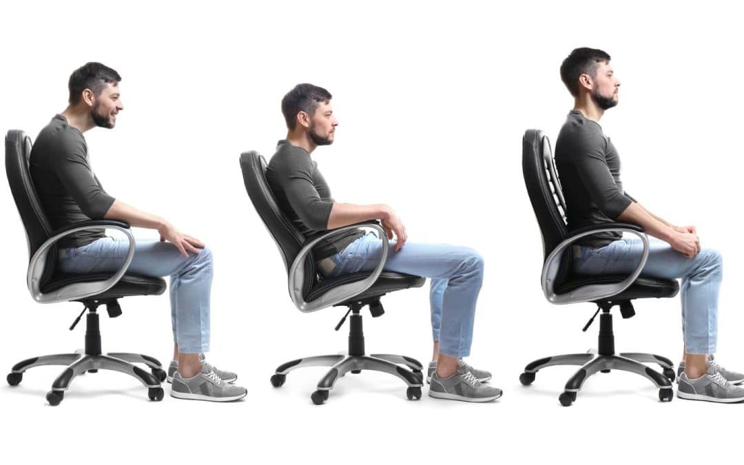
Good posture is more than just standing up straight; it’s a vital aspect of your overall health that impacts everything from your musculoskeletal system to your mental well-being. Dynamic posture ensures that your body functions efficiently, while postural stagnancy can contribute to chronic pain, decreased range of motion, and other health issues. In today’s digital world, where many of us spend hours sitting or hunching over devices, understanding the importance of posture is essential.
This blog post delves into how posture affects your health, the consequences of poor posture, and how kinesiology—a field closely related to physiotherapy—can help you correct postural imbalances and improve overall well-being.
What Is Posture?
Posture refers to the alignment of the body’s muscles and skeleton when you sit, stand, or lie down. Good posture maintains the natural curves of the spine, minimizes stress on the musculoskeletal system, and promotes efficient movement patterns. Key characteristics of good posture include:
- A neutral spine
- Shoulders aligned with the hips
- Even weight distribution
- A balanced head position
Why Good Posture Matters
Good posture is essential for overall health. Here’s why:
- Musculoskeletal Health: dynamic posture ensures that bones and joints are in alignment and exposed to a variety of movement opportunities. This allows the brain to assess environmental safety ( reducing the likelihood of pain) and promoting physiological effects such as joint lubrication and parasympathetic regulation. These in turn, reduce the risk of musculoskeletal issues such as back pain, joint pain, and muscular imbalances.
- Chronic Pain Prevention: Poor posture often leads to chronic pain, especially in the lower back, neck, and shoulders. By maintaining good posture, you can prevent or alleviate many forms of chronic pain.
- Efficient Movement Patterns: Good posture promotes efficient movement patterns, reducing the strain on muscles and ligaments and enhancing overall physical performance.
- Respiratory Function: A hunched posture can compress the diaphragm and restrict lung capacity, leading to shallow breathing and reduced oxygen intake. Good posture allows for optimal lung function and better breathing.
- Circulation and Digestion: Proper posture ensures that blood circulates efficiently throughout the body and that internal organs function properly. Poor posture can compress abdominal organs, leading to digestive issues.
- Mental Well-being: Studies suggest that posture can influence mood and mental health and vice versa. An upright posture has been linked to improved self-esteem and reduced stress, while slouching is associated with negative emotions.
The Consequences of Poor Posture
In today’s sedentary lifestyle, poor posture has become increasingly common, leading to various health issues:
- Back Pain and Other Musculoskeletal Problems: Poor posture is a leading cause of lower back pain and other musculoskeletal problems. Over time, it can cause the spine to become sensitive and painful. This in combination with a lifestyle low in general fitness can contribute to conditions such as sciatica and herniated discs.
- Muscular Imbalances: Poor posture can cause certain muscles to become overactive while others weaken, resulting in muscular imbalances that affect overall movement and posture.
- Limited Range of Motion: Postural imbalances can lead to tight muscles and restricted joint movement, reducing your range of motion and making everyday activities more challenging.
- Increased Risk of Injury: Poor posture can lead to improper movement patterns, increasing the risk of injuries, especially during physical activities.
- Fatigue: Poor posture forces the body to work harder to maintain balance, leading to increased fatigue and decreased energy levels.
How Kinesiology Can Help Correct and Improve Posture
What Is Kinesiology?
Kinesiology is the scientific study of human movement, encompassing various disciplines such as biomechanics, anatomy, and physiology. It’s closely related to physiotherapy and focuses on assessing and correcting movement patterns to improve overall health. Kinesiologists use a holistic approach to identify postural issues, address muscular imbalances, and develop customized exercise programs to correct poor posture.
The Role of Kinesiology in Posture Correction
- Postural Assessment: Kinesiologists perform detailed postural assessments to identify areas of imbalance and weakness. This may include observing how you stand, sit, and move, as well as testing muscle strength and flexibility.
- Customized Exercise Programs: Based on the assessment, a kinesiologist will develop a personalized exercise program aimed at correcting postural imbalances. This program may include stretching tight muscles, strengthening weak ones, and improving overall range of motion.
- Ergonomic Advice: Kinesiologists often provide advice on ergonomics, helping you set up your workspace or living environment to promote good posture. This might include recommendations for chair height, monitor placement, and proper sitting posture.
- Manual Therapy: Some kinesiologists use manual therapy techniques, such as massage or joint mobilization, to relieve tension, improve joint mobility, and support better posture.
- Ongoing Monitoring and Support: Posture correction is an ongoing process. Kinesiologists provide continuous support and adjustments to your exercise program, ensuring you make steady progress toward better posture and overall health.
The Science Behind Kinesiology and Posture Correction
Several studies highlight the effectiveness of kinesiology-based interventions for improving posture and alleviating chronic pain:
- Musculoskeletal Improvements: Research published in the Journal of Back and Musculoskeletal Rehabilitation found that participants in a kinesiology-based program experienced significant improvements in posture and a reduction in chronic back pain.
- Enhanced Range of Motion: A study in the Journal of Physical Therapy Science demonstrated that kinesiology-based exercises improved the range of motion and reduced muscular imbalances in individuals with forward head posture.
- Chronic Pain Relief: According to the Journal of Orthopaedic & Sports Physical Therapy, kinesiology-based interventions significantly reduced chronic pain in participants by correcting postural imbalances and improving movement patterns.
- Improved Respiratory Function: Research in the International Journal of Osteopathic Medicine showed that postural correction through kinesiology improved respiratory function, particularly in individuals with a hunched posture.
Practical Tips for Maintaining Good Posture
In addition to working with a kinesiologist, there are several steps you can take to improve and maintain good posture:
- Be Mindful of Your Posture: Regularly check your posture throughout the day, whether you’re sitting, standing, or moving. Make small adjustments to offer the body a variety of movement options.
- Ergonomics: Set up your workspace to promote good posture. Ensure that your chair, desk, and computer are at the right height to keep your spine in a neutral position.
- Strengthen Your Core: A strong core is essential for maintaining good posture. Include exercises that target the abdominal muscles, lower back, and hips in your routine.
- Stretch Regularly: Stretching can help relieve muscle tightness and improve flexibility, making it easier to maintain good posture. Focus on stretching the chest, shoulders, and hip flexors.
- Stay Active: Regular physical activity helps maintain muscle balance and flexibility, which are crucial for good posture. Incorporate a variety of activities, including strength training, cardio, and flexibility exercises.
- Use a Supportive Mattress and Pillow: Your sleeping posture is just as important as your waking posture. Choose a mattress and pillow that support the natural curves of your spine.
Conclusion
Posture plays a crucial role in your overall health, influencing everything from musculoskeletal function to mental well-being. Poor posture can lead to chronic pain, reduced mobility, and impaired bodily functions, but the good news is that it can be corrected. Kinesiology offers a comprehensive approach to assessing, correcting, and improving posture, helping you to address the root causes of poor posture and achieve better health.
Whether you’re experiencing chronic pain, recovering from an injury, or simply looking to improve your posture for long-term health benefits, working with a kinesiologist like those at South Island Physiotherapy can provide you with the tools and support you need to achieve good posture and overall well-being.

by Jason Nenzel | Jul 19, 2024 | news
Effects, Mechanisms, and Supporting Evidence of Myofascial Release Therapy
Myofascial release therapy (MFR) is a type of manual therapy that focuses on relieving tension in the fascia, the connective tissue that surrounds and supports muscles and other structures throughout your body. This therapy may be beneficial for various conditions, particularly those involving muscle and joint pain. Let’s explore the principles of myofascial release therapy, its proposed physiological effects, and the evidence supporting its use.
What is Myofascial Release Therapy?
Myofascial release therapy involves the application of sustained pressure and gentle stretching to the myofascial tissue with the aim of releasing restrictions and tension. This hands-on technique is typically performed by physical therapists, registered massage therapists, and occupational therapists. During therapy sessions, the therapist will massage and stretch the fascia, targeting areas that feel stiff and tight, to improve the elasticity and mobility of the tissue.
Proposed Physiological Effects of Myofascial Release Therapy
1. Reduction of Fascial Restrictions
- Theory: Fascia can become restricted due to trauma, inflammation, or poor posture, leading to decreased mobility and pain. Myofascial release therapy focuses on releasing these restrictions to restore normal function.
- Evidence: Some studies have shown that MFR can increase tissue elasticity and reduce fascial stiffness, which may help improve range of motion and alleviate pain.
2. Pain Relief
- Theory: By releasing fascial restrictions and improving blood flow, myofascial release therapy can reduce pain and discomfort associated with various musculoskeletal conditions.
- Evidence: Some research indicates that MFR can be effective in reducing pain in conditions such as chronic low back pain, fibromyalgia, and plantar fasciitis.
3. Improved Circulation
- Theory: MFR is thought to enhance blood flow to affected areas, promoting healing and reducing inflammation.
- Evidence: Some studies suggest that MFR can improve microcirculation and lymphatic flow, aiding in the removal of metabolic waste products and reducing inflammation.
4. Enhanced Muscle Function
- Theory: Releasing fascial tension can improve muscle function by allowing muscles to move more freely and efficiently.
- Evidence: Evidence supports the idea that MFR can improve muscle activation and coordination, potentially enhancing athletic performance and reducing the risk of injury.
5. Stress Reduction
- Theory: The gentle, sustained pressure of MFR can activate the parasympathetic nervous system, promoting relaxation and reducing stress.
- Evidence: Preliminary research suggests that MFR may have beneficial effects on stress reduction and overall mental well-being.
Evidence Supporting Myofascial Release Therapy
While anecdotal reports and clinical experience have long supported the use of MFR, scientific research has begun to provide more rigorous evidence of its effectiveness. Here are some key findings:
- Chronic Low Back Pain: A systematic review and meta-analysis found that MFR significantly reduces pain and improves functional outcomes in patients with chronic low back pain.
- Fibromyalgia: Studies have shown that MFR can reduce pain, improve sleep quality, and enhance the quality of life in patients with fibromyalgia.
- Plantar Fasciitis: Research indicates that MFR can be an effective treatment for reducing pain and improving function in individuals with plantar fasciitis.
- Carpal Tunnel Syndrome: MFR may help reduce pain and improve hand function in patients with carpal tunnel syndrome.
How Myofascial Release Therapy Works
- Assessment: The therapist assesses the patient’s posture, movement patterns, and areas of pain or restriction. This may involve identifying myofascial trigger points, which are stiff areas in the muscle that cause pain.
- Application of Pressure: The therapist applies gentle, sustained pressure to specific areas of the fascia using their hands, elbows, or specialized tools like foam rollers.
- Stretching and Movement: The therapist may incorporate gentle stretching and movement to help release fascial restrictions.
- Monitoring Response: The therapist monitors the patient’s response to the treatment and adjusts the pressure and techniques as needed.
Self-Myofascial Release
Self-myofascial release involves using tools like foam rollers or roller massagers to apply pressure to the fascia. This can be done at home and is a convenient way to manage pain and maintain flexibility between therapy sessions. Self-myofascial release might involve rolling the foam roller over the muscles and holding pressure on tight spots for 30-60 seconds.
Conditions Treated by Myofascial Release
Myofascial release therapy can help with various conditions, including:
- Myofascial Pain Syndrome: A chronic pain disorder caused by sensitivity and tightness in your myofascial tissues. Pain originates from specific trigger points and can be widespread.
- Low Back Pain: MFR can help alleviate chronic low back pain by releasing tight fascia in the lumbar region.
- Fibromyalgia: MFR may reduce widespread pain and improve quality of life for fibromyalgia patients.
- Carpal Tunnel Syndrome: MFR can relieve pain and improve hand function by targeting the fascia in the wrists and hands.
- Plantar Fasciitis: MFR can reduce pain in the feet by releasing tight fascia in the plantar area.
Benefits of Myofascial Release Therapy
- Pain Relief: MFR can provide significant pain relief for various musculoskeletal conditions.
- Improved Range of Motion: By releasing fascial restrictions, MFR can enhance flexibility and mobility.
- Reduced Stress: The relaxation response elicited by MFR can help reduce overall stress levels.
- Enhanced Muscle Function: Improved fascial mobility can lead to better muscle function and coordination.
Conclusion
The specific physiological changes that may occur during Myofascial release therapy remain a hot topic for debate however it maintains itself as a popular and promising modality for addressing a range of musculoskeletal conditions and improving overall physical function ( at least in the short term). Whether performed by a physical therapist or through self-myofascial release techniques, this type of therapy may benefit those experiencing pain and discomfort from various conditions. More research is needed to fully understand its mechanisms and long-term effects. While current evidence supports its use for pain relief, improved mobility, and enhanced muscle function, It also remains true that progressive loading of the injured region is the most predictable form of intervention to produce durable long-term changes in physiological tissue. As we often state in the clinic, the body is an ecosystem and responds well to active stressors. Passive stressors like MFR are a wonderful adjunct to active exercise in order to facilitate confidence and comfort while pursuing long term results.
If you’re considering myofascial release therapy, consult with a qualified healthcare professionals like the ones at South Island Physiotherapy to determine if it’s appropriate for your specific needs and conditions. By incorporating myofascial release into your wellness routine, you may experience significant improvements in your physical health and overall well-being.
References
- Ajimsha, M. S., Al-Mudahka, N. R., & Al-Madzhar, J. A. (2015). Effectiveness of myofascial release: Systematic review of randomized controlled trials. Journal of Bodywork and Movement Therapies, 19(1), 102-112.
- Kim, J. H., Kim, S. H., & Kim, Y. H. (2014). The effect of myofascial release on pain and functional outcomes in patients with chronic low back pain: A meta-analysis. Journal of Physical Therapy Science, 26(1), 177-179.
- Castro-Sánchez, A. M., Matarán-Peñarrocha, G. A., Arroyo-Morales, M., et al. (2011). Effects of myofascial release techniques on pain and sleep quality in patients with fibromyalgia: A randomized controlled trial. Journal of Manipulative and Physiological Therapeutics, 34(10), 507-513.
- Hanten, W. P., Olson, S. L., Butts, N. L., & Nowicki, A. L. (2000). Effectiveness of a home program of ischemic pressure followed by sustained stretch for treatment of myofascial trigger points. Physical Therapy, 80(10), 997-1003.
- Beardsley, C., & Škarabot, J. (2015). Effects of self-myofascial release: A systematic review. Journal of Bodywork and Movement Therapies, 19(4), 747-758.
- Barnhart, A., Davenport, T. E., & Chinn, L. A. (2017). The effects of myofascial release on the autonomic nervous system response. Journal of Bodywork and Movement Therapies, 21(1), 5-11.
by Jason Nenzel | Jun 10, 2024 | news
Health Consequences, Benefits of Exercise, and When to Seek Medical Attention
Blood pressure is a critical health indicator often referred to as the silent killer due to its subtle yet potentially devastating effects. This blog post educates on high blood pressure, the health consequences of hypertension, the positive effects of exercise on blood pressure, and when to seek medical attention.
What is Blood Pressure?
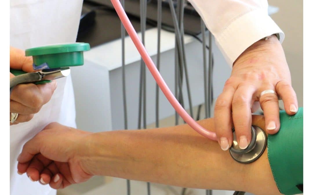
Blood pressure is the force exerted by circulating blood against the walls of the arteries. It is measured using a blood pressure monitor and recorded as two numbers:
- Systolic pressure: The top number represents the pressure in your arteries when your heart beats.
- Diastolic pressure: The bottom number indicates the pressure in your arteries when your heart rests between beats.
A normal blood pressure reading is typically around 120/80 mmHg, according to the American Heart Association. Blood pressure can fluctuate based on activity, stress, diet, and overall health.
Health Consequences of High Blood Pressure
High blood pressure, or hypertension, occurs when the force of the blood against the artery walls is consistently too high. This condition can lead to severe health problems, including:
1. Heart Disease and Stroke
Hypertension increases the risk of heart disease, including heart attacks and strokes. The increased pressure can damage the arteries, making them less elastic, which decreases the flow of blood and oxygen to the heart and brain.
2. Aneurysm
Persistent high blood pressure can cause blood vessels to weaken and bulge, forming an aneurysm. If an aneurysm ruptures, it can be life-threatening.
3. Heart Failure
The heart has to work harder to pump blood against the higher pressure in the vessels, leading to thickening of the heart muscle. Over time, this can cause the heart to struggle to pump enough blood to meet the body’s needs, leading to heart failure.
4. Kidney Damage
Hypertension can damage the blood vessels in the kidneys, affecting their ability to filter waste from the blood effectively. This can lead to kidney disease or failure.
5. Vision Loss
High blood pressure can damage the tiny, delicate blood vessels that supply blood to the eyes, leading to vision problems or blindness.
6. Metabolic Syndrome
This syndrome involves a combination of disorders, including high blood pressure, high blood sugar, excess body fat around the waist, and abnormal cholesterol levels. It increases the risk of heart disease, stroke, and diabetes.
Positive Effects of Exercise on Blood Pressure
Regular physical activity is one of the most effective ways to prevent or manage hypertension. Here’s how exercise can positively impact blood pressure:
1. Lowers Blood Pressure
Exercise helps lower blood pressure by improving the heart’s efficiency, allowing it to pump blood with less effort, reducing the force on the arteries.
2. Promotes Weight Loss
Maintaining a healthy weight is crucial for blood pressure control. Exercise helps burn calories and reduces body fat, which can help lower blood pressure.
3. Improves Heart Health
Regular physical activity strengthens the heart muscle, improving its ability to pump blood and reducing the workload on the arteries.
4. Reduces Stress
Exercise can lower stress levels, which can contribute to high blood pressure. Activities like walking, swimming, and yoga can help promote relaxation and reduce stress hormones.
5. Improves Sleep
Regular physical activity can improve sleep quality, which is important for maintaining healthy blood pressure levels.
Recommended Exercises for Blood Pressure Management
- Aerobic exercises: Walking, jogging, cycling, swimming, and dancing.
- Strength training: Lifting weights or using resistance bands.
- Flexibility and balance exercises: Yoga and tai chi.
Exercise Guidelines
- Aim for at least 150 minutes of moderate-intensity aerobic activity or 75 minutes of vigorous-intensity activity each week.
- Include muscle-strengthening activities on two or more days a week.
- Start slowly and gradually increase the intensity and duration of your workouts.
Measuring Blood Pressure at Home
Monitoring blood pressure at home is a practical way to keep track of your health. Using a home blood pressure monitor allows you to regularly measure your blood pressure and understand how lifestyle changes impact your health.
Steps to Measure Your Blood Pressure at Home:
- Choose a Home Blood Pressure Monitor: Select a reliable device, preferably one validated by the American Heart Association.
- Prepare for Measurement: Sit quietly for five minutes before measuring. Avoid caffeine, exercise, and smoking 30 minutes prior.
- Position Correctly: Sit with your back straight and supported, feet flat on the floor, and arm supported at heart level.
- Take Multiple Readings: Take two or three readings one minute apart and record the results.
When to Seek Medical Attention
It’s important to regularly measure your blood pressure and seek medical attention if you experience any of the following:
1. Consistently High Readings
If your blood pressure readings are consistently above 140/90 mmHg, it’s time to consult a healthcare provider.
2. Symptoms of Severe Hypertension
Symptoms such as severe headaches, shortness of breath, nosebleeds, chest pain, visual changes, or blood in the urine require immediate medical attention.
3. Medication Side Effects
If you’re experiencing side effects from blood pressure medication, consult your doctor to adjust the dosage or explore alternative treatments.
4. Uncontrolled Blood Pressure
Despite lifestyle changes and medication, if your blood pressure remains high, further medical evaluation and intervention may be necessary.
5. Other Health Conditions
If you have conditions like diabetes, kidney disease, or heart disease, regular blood pressure monitoring and management are crucial.
Conclusion
Understanding and managing blood pressure is vital for maintaining overall health and preventing serious health issues. Regular exercise, a healthy diet, stress management, and regular blood pressure checks are key components of blood pressure management. If you experience any concerning symptoms or have consistently high readings, seek medical attention promptly to ensure proper care and intervention. Your heart and arteries will thank you for it!
by Jason Nenzel | May 20, 2024 | news
Diagnostic Technologies and Their Clinical Indications in Musculoskeletal Care
Medical imaging has transformed modern healthcare, providing critical insights that enable accurate diagnosis and effective treatment of many pathologies, including musculoskeletal injuries. Each imaging modality employs unique technologies and serves specific clinical purposes.
This guide delves into the primary types of medical imaging used to assist care of acute and chronic injuries, their underlying technologies, and their common clinical indications, highlighting the role of imaging in enhancing diagnostic accuracy and patient care.
1. X-ray Imaging
Technology:
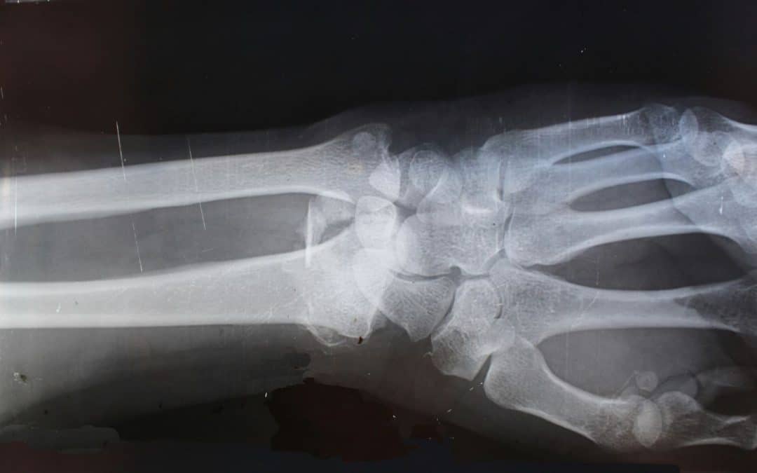
X-ray imaging is one of the oldest and most widely used imaging techniques. It uses ionizing radiation to produce images of the body’s internal structures. An X-ray machine emits X-ray beams that pass through the body and are captured by a detector on the other side. The varying absorption rates of different tissues create a contrast image, with bones appearing white, soft tissues in shades of gray, and air spaces black.
Clinical Indications:
X-rays are extensively used in diagnosing and managing a wide array of musculoskeletal conditions. Here are some of their primary applications:
Fracture Detection and Management:
- Acute Fractures: X-rays are the first-line imaging modality for detecting acute fractures. They can identify the location, type, and extent of bone breaks, guiding initial treatment and management.
- Stress Fractures: While early-stage stress fractures might not be visible on initial X-rays, they can show up on follow-up X-rays as callus formation or periosteal reaction.
- Pediatric Fractures: X-rays are crucial for evaluating fractures in children, including growth plate (physeal) injuries, which require careful management to avoid growth disturbances.
Joint Pathologies:
- Arthritis: X-rays are instrumental in diagnosing various types of arthritis. They can show joint space narrowing, osteophyte formation, subchondral sclerosis, and other characteristic changes associated with osteoarthritis, rheumatoid arthritis, and other arthritic conditions.
- Joint Dislocations: X-rays provide clear images of joint dislocations, helping in the assessment of the extent of displacement and guiding reduction procedures.
Bone Pathologies:
- Bone Tumors: X-rays can identify primary bone tumors and metastatic lesions. They help in characterizing bone lesions based on their appearance, such as lytic or sclerotic patterns.
- Osteomyelitis: X-rays can detect signs of bone infection, including periosteal elevation, bone destruction, and new bone formation.
Spinal Disorders:
- Degenerative Changes: X-rays of the spine are used to assess degenerative changes, such as disc space narrowing, osteophytes, and facet joint arthritis.
- Scoliosis: X-rays provide a clear assessment of spinal curvature in scoliosis, helping in monitoring the progression and planning treatment.
Soft Tissue Assessment:
- Calcifications: X-rays can detect soft tissue calcifications, such as myositis ossificans or calcific tendinitis.
- Foreign Bodies: X-rays are useful for locating radiopaque foreign bodies in soft tissues, aiding in their removal.
Preoperative Planning and Postoperative Evaluation:
- Preoperative Planning: X-rays provide essential anatomical details needed for planning orthopedic surgeries, such as fracture fixation, joint replacement, and spinal fusion.
- Postoperative Assessment: X-rays are used to evaluate the positioning and integration of surgical implants, healing of fractures, and detection of potential complications like non-union or hardware failure.
Advances:
Digital X-ray technology has significantly improved image quality and reduced radiation exposure compared to traditional film X-rays. Additionally, portable X-ray machines have made it possible to perform imaging procedures at the bedside, enhancing accessibility in emergency and critical care settings.
2. Computed Tomography (CT)
Technology:
Computed Tomography (CT) scanning combines X-ray equipment with advanced computer processing to create detailed cross-sectional images of the body. During a CT scan, the X-ray tube rotates around the patient, capturing multiple images from different angles. These images are then processed by a computer to produce cross-sectional slices, which can be further reconstructed into 3D images.
Clinical Indications:
CT scans are extensively used in diagnosing and managing a wide range of musculoskeletal conditions. Here are some of its primary applications:
Fracture Detection and Assessment:
- Complex Fractures: CT is invaluable in evaluating complex fractures, particularly in areas with intricate anatomy, such as the pelvis, spine, and facial bones. It provides detailed information on fracture lines, displacement, and comminution.
- Subtle Fractures: CT can detect fractures that may not be visible on conventional X-rays, such as stress fractures and small cortical breaks.
Bone and Joint Pathologies:
- Arthritis: CT imaging is used to assess the extent of joint damage in osteoarthritis and other arthritic conditions, visualizing bone spurs, joint space narrowing, and subchondral cysts.
- Bone Tumors: CT scans help in the characterization and staging of bone tumors, providing detailed information on the lesion’s size, location, and potential cortical involvement.
- Osteomyelitis: CT is useful in detecting bone infections, revealing areas of bone destruction, periosteal reaction, and abscess formation.
Spinal Disorders:
- Disc Herniations: CT myelography, which involves the injection of contrast material into the spinal canal, enhances the visualization of disc herniations and their effect on nerve roots and the spinal cord.
- Degenerative Changes: CT is effective in assessing degenerative spinal conditions such as spondylosis, facet joint arthritis, and spinal stenosis, providing detailed images of bony changes and foraminal narrowing.
- Trauma: In cases of spinal trauma, CT quickly identifies fractures, dislocations, and bone fragments, guiding immediate management and surgical intervention if necessary.
Preoperative Planning and Postoperative Evaluation:
- Surgical Planning: CT provides precise anatomical details crucial for planning orthopedic surgeries, such as fracture fixation, joint replacement, and spinal fusion. 3D reconstructions are particularly valuable in visualizing complex deformities and planning corrective procedures.
- Postoperative Assessment: CT scans are used to evaluate the position and integrity of surgical implants, detect postoperative complications, and monitor the healing process.
Assessment of Bone Density and Structure:
- Osteoporosis: Quantitative CT (QCT) measures bone mineral density, aiding in the diagnosis and management of osteoporosis. QCT provides volumetric measurements of bone density, which are more accurate than conventional dual-energy X-ray absorptiometry (DEXA) scans in some cases.
Vascular Evaluation:
- Vascular Imaging: CT angiography (CTA) evaluates blood vessels, identifying conditions such as aneurysms, vascular malformations, and arterial stenosis. In the context of musculoskeletal imaging, CTA can assess vascular injuries associated with fractures or dislocations.
Advances:
Modern CT scanners offer high-speed imaging and lower doses of radiation through techniques like helical (spiral) CT and dual-energy CT. These advancements improve diagnostic accuracy and patient safety by minimizing radiation exposure.
3. Magnetic Resonance Imaging (MRI)
Technology:
Magnetic Resonance Imaging (MRI) uses powerful magnets, radio waves, and a computer to produce detailed images of the body’s organs and tissues. In an MRI scan, the magnetic field temporarily aligns hydrogen atoms in the body. Radiofrequency pulses then disrupt this alignment, and the returning signals are used to generate images. MRI provides excellent soft tissue contrast without using ionizing radiation.
Clinical Indications:
Joint Pathologies:
- Cartilage Lesions: MRI is the gold standard for evaluating cartilage integrity and detecting chondral lesions and osteochondritis dissecans. High-resolution imaging allows for detailed assessment of cartilage thickness and surface irregularities.
- Meniscal Tears: In the knee, MRI is particularly useful for diagnosing meniscal tears, providing detailed images of the menisci and surrounding structures.
- Labral Tears: MRI arthrography, which involves injecting contrast material into the joint, enhances the visualization of the labrum in the shoulder and hip, aiding in the diagnosis of labral tears and impingement syndromes.
Tendon and Ligament Injuries:
- Rotator Cuff Tears: MRI accurately detects partial and complete tears of the rotator cuff tendons in the shoulder. It also assesses the extent of tendon retraction and muscle atrophy, guiding surgical planning.
- Anterior Cruciate Ligament (ACL) Tears: MRI is essential for diagnosing ACL injuries in the knee, visualizing the ligament’s integrity and associated injuries to other structures like the menisci and collateral ligaments.
- Achilles Tendon Injuries: MRI evaluates the Achilles tendon for tears, tendinopathy, and associated conditions such as retrocalcaneal bursitis.
Bone and Marrow Pathologies:
- Stress Fractures: MRI is more sensitive than X-ray in detecting early stress fractures and bone marrow edema, providing critical information for early intervention and management.
- Bone Tumors and Infections: MRI is highly effective in characterizing bone tumors and infections (osteomyelitis), offering detailed images of bone marrow changes, tumor extent, and soft tissue involvement.
Muscle Injuries and Disorders:
- Muscle Tears: MRI accurately identifies muscle strains and tears, grading the severity of the injury and helping guide rehabilitation strategies.
- Myopathies: MRI can detect inflammatory and metabolic myopathies, visualizing muscle edema, fatty infiltration, and atrophy.
Nerve Disorders:
- Peripheral Neuropathies: MRI can visualize peripheral nerves and diagnose compressive neuropathies, such as carpal tunnel syndrome and ulnar nerve entrapment. It helps identify the site and cause of nerve compression.
- Brachial Plexus Injuries: MRI is crucial in evaluating traumatic and non-traumatic brachial plexus injuries, providing detailed images of nerve roots, trunks, and associated lesions.
Spine Disorders:
- Disc Herniations: MRI is the preferred imaging modality for diagnosing intervertebral disc herniations, visualizing the extent of disc protrusion and its impact on adjacent neural structures.
- Spinal Stenosis: MRI assesses spinal canal narrowing and nerve root compression, aiding in the diagnosis and management of spinal stenosis.
- Vertebral Infections and Tumors: MRI provides detailed images of vertebral bodies and intervertebral discs, essential for diagnosing infections (spondylodiscitis) and tumors.
- Neurological Disorders: MRI is the gold standard for diagnosing brain tumors, strokes, multiple sclerosis, and spinal cord injuries. It provides high-resolution images of brain and spinal cord structures.
- Musculoskeletal Problems: MRI is ideal for evaluating joint abnormalities, soft tissue injuries, and spinal disc issues. It can detect ligament tears, cartilage damage, and other musculoskeletal conditions.
- Cardiac Imaging: Cardiac MRI assesses heart structure and function, detecting conditions such as cardiomyopathy, congenital heart disease, and myocardial infarction ( heart attack).
Advances:
Functional MRI (fMRI) measures brain activity by detecting changes in blood flow, providing insights into brain function and aiding in pre-surgical planning. Additionally, advancements in MRI technology, such as higher field strengths (3T and 7T MRI), enhance image resolution and diagnostic capabilities.
4. Ultrasound
Technology:
Ultrasound imaging uses high-frequency sound waves to create real-time images of the inside of the body. A transducer emits sound waves and records the echoes as they bounce back from internal tissues. The captured echoes are used to construct images, which can be viewed in real-time, making ultrasound particularly useful for dynamic studies.
Clinical Indications:
Tendon and Ligament Injuries:
- Tendon Tears and Tendinopathy: Ultrasound is highly effective in detecting partial and complete tendon tears, as well as tendinopathies (degenerative changes in tendons). Common sites include the rotator cuff in the shoulder, Achilles tendon, and patellar tendon.
- Ligament Injuries: Ultrasound can identify ligament sprains and tears, particularly in the ankle, knee, and wrist. Dynamic imaging can assess the stability of ligaments during stress maneuvers.
Muscle Injuries:
- Muscle Tears: Acute muscle injuries, such as strains and tears, can be readily identified. Ultrasound helps in grading the severity of muscle injuries, guiding appropriate treatment and rehabilitation.
- Muscle Hernias: The real-time capabilities of ultrasound are beneficial in diagnosing muscle hernias, where a portion of the muscle protrudes through a defect in the fascia.
Joint Pathologies:
- Joint Effusions: Ultrasound can detect fluid accumulation within joints, indicative of inflammation, infection, or injury. It also assists in guiding joint aspiration procedures to remove fluid for diagnostic and therapeutic purposes.
- Arthritis: Inflammatory arthritis, such as rheumatoid arthritis, can be monitored using ultrasound to assess synovial thickening, joint effusions, and erosions.
Nerve Entrapments:
- Carpal Tunnel Syndrome: Ultrasound is useful in diagnosing compressive neuropathies like carpal tunnel syndrome, where the median nerve is compressed at the wrist. It visualizes nerve swelling and structural changes.
- Other Entrapments: Conditions such as ulnar nerve entrapment at the elbow and tarsal tunnel syndrome in the ankle can also be evaluated.
Bursitis and Cystic Lesions:
- Bursitis: Ultrasound identifies inflammation of bursae, such as subacromial bursitis in the shoulder and trochanteric bursitis in the hip.
- Cysts: Ganglion cysts, Baker’s cysts, and other fluid-filled lesions can be accurately detected and characterized.
Guided Interventions:
- Injections and Aspirations: Ultrasound guidance improves the accuracy of therapeutic injections (e.g., corticosteroids) and aspirations (e.g., fluid removal) into joints, tendons, and soft tissue structures. This enhances the efficacy and safety of these procedures.
- Biopsies: Ultrasound guidance is also used for performing needle biopsies of soft tissue masses to obtain tissue samples for pathological analysis
Advances:
Doppler ultrasound measures blood flow through vessels, aiding in the diagnosis of blockages, clots, and other vascular conditions. Advances in 3D and 4D ultrasound provide more detailed and dynamic images, improving diagnostic accuracy in various clinical scenarios.
The Role of Imaging in Enhancing Diagnostic Accuracy
Medical imaging is integral to modern diagnostics, offering a non-invasive means to visualize internal structures and functions. The different imaging modalities—X-ray, CT, MRI, ultrasound, and nuclear medicine—each have distinct strengths and clinical indications. Selecting the appropriate imaging technique based on the clinical scenario ensures optimal diagnostic accuracy and patient care.
Importance of Image Data and Medical Imaging Equipment
High-quality image data is crucial for accurate diagnosis and treatment planning. Advanced imaging equipment, including digital X-ray machines, high-resolution CT scanners, and high-field MRI systems, enhances the quality of images and diagnostic capabilities. Continuous advancements in imaging technology contribute to improved patient outcomes and more precise medical interventions.
Conclusion and a Word of Caution
Medical imaging has transformed healthcare by providing detailed insights into the human body, facilitating accurate diagnosis and effective treatment. Understanding the technologies and clinical indications for various imaging modalities enables healthcare professionals to choose the most appropriate methods for their patients. Continued advancements in imaging technology promise even greater precision, reduced radiation exposure, and improved patient outcomes.
With that said, diagnostic imaging does not show pain. In the world of conservative care, imaging rarely changes the course of treatment unless there is a concern for a more profound, high-risk injury ( like cancer or fracture) or evidence-informed conservative care has failed to assist in the resolution of the condition.
There is a host of evidence (that could be a blog post in and of itself) on how premature imaging of a biologically safe pain experience can lead to prolonged pain and even push someone into chronic disability, so these technologies need to be used practically and judiciously if we are being truly patient centred with our approach to injury. In short, an image does not trump the patient’s pain experience. It serves as a valuable tool to expand the diagnostic narrative when clinically indicated.
We at South Island Physiotherapy hope this review of common musculoskeletal medical imaging techniques provides insight into why certain types of imaging may be prescribed for your condition, and we are here to help you make sense of how they can assist you in your recovery.

